. 2010 Oct;21(10):1691-701.
doi: 10.1681/ASN.2010030295. Epub 2010 Aug 5.

Glomerular structure and function require paracrine, not autocrine, VEGF-VEGFR-2 signaling

Affiliations

Glomerular structure and function require paracrine, not autocrine, VEGF-VEGFR-2 signaling

Karen Sison et al. J Am Soc Nephrol. 2010 Oct.

Erratum in

  • J Am Soc Nephrol. 2011 Jul;22(7):1390

Abstract

VEGF is a potent vascular growth factor produced by podocytes in the developing and mature glomerulus. Specific deletion of VEGF from podocytes causes glomerular abnormalities including profound endothelial cell injury, suggesting that paracrine signaling is critical for maintaining the glomerular filtration barrier (GFB). However, it is not clear whether normal GFB function also requires autocrine VEGF signaling in podocytes. In this study, we sought to determine whether an autocrine VEGF-VEGFR-2 loop in podocytes contributes to the maintenance of the GFB in vivo. We found that induced, whole-body deletion of VEGFR-2 caused marked abnormalities in the kidney and also other tissues, including the heart and liver. By contrast, podocyte-specific deletion of the VEGFR-2 receptor had no effect on glomerular development or function even up to 6 months old. Unlike cell culture models, enhanced expression of VEGF by podocytes in vivo caused foot process fusion and alterations in slit diaphragm-associated proteins; however, inhibition of VEGFR-2 could not rescue this defect. Although VEGFR-2 was dispensable in the podocyte, glomerular endothelial cells depended on VEGFR-2 expression: postnatal deletion of the receptor resulted in global defects in the glomerular microvasculature. Taken together, our results provide strong evidence for dominant actions of a paracrine VEGF-VEGFR-2 signaling loop both in the developing and in the filtering glomerulus. VEGF produced by the podocyte regulates the structure and function of the adjacent endothelial cell.


Figures

Figure 1.
Figure 1.
Whole body deletion of VEGFR-2 results in vascular defects, with glomerular thrombotic microangiopathy. (A) Conditional whole body VEGFR-2 knockout mice contain five transgenes. rtTA is expressed by all cells of the body and, with DOX, binds to the tetO-regulated Cre protein. Cre induces recombination between the loxP sites on the VEGFR-2 allele and results in VEGFR-2 deletion in all cells. (B) PCR analysis of tail genomic DNA shows Cre-mediated excision of the floxed VEGFR-2 region (floxed allele, 439 bp; wild-type allele, 322 bp; deleted allele, 218 bp). (C) Light micrographs show pristine glomeruli in control mice (i through iii). Glomeruli from mutant mice show global glomerular damage [(iv through ix); black arrows in (iv)]. Features of chronic TMA include fragmented red blood cells [black arrows in (vi)], interposition of mesangial cells [white arrowhead in (viii)] and marked thickening of the wall of a capillary loop [black arrowhead in (ix)]. Top row and (viii): periodic acid–Schiff stain; (ii and v): hematoxylin and eosin stain. Bottom row: Trichrome masson stain. Original magnifications: ×100 (i and iv); ×400 (ii and v); ×800 (iii, vi, and ix); ×1000 (viii).
Figure 2.
Figure 2.
Podocytes do not express VEGFR-2 in vivo. (A) GFP is knocked into the VEGFR-2 locus. GFP staining shows that VEGFR-2 is expressed only in endothelial cells in the glomerulus (arrows). (B) FACS sorting of primary podocytes. Glomeruli were isolated from Nephrin-CFP transgenic mice which contain a CFP reporter gene that is driven by the podocyte-specific nephrin promoter. Glomerular cells were dissociated enzymatically and were sorted into two fractions on the basis of fluorescence, CFP-expressing (CFP+) primary podocytes and CFP-negative (CFP) endothelial and mesangial cells. RNA were extracted from each fraction and converted into cDNA. (C and D) VEGF receptor mRNA expression in FACS-sorted cells was assessed by reverse transcriptase real-time PCR (CFP+ = podocytes, CFP = endothelial cells and mesangial cells, GFP+ = endothelial cells, GFP = podocytes, mesangial cells). Values are expressed as fold increase over CFP, CFP+, or GFP+ cell fraction and represent mean ± SEM (n = 3 to 4 in all groups). t test was used for statistical analyses (*P < 0.05, **P < 0.01, ***P < 0.001).
Figure 3.
Figure 3.
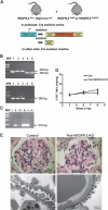
Genetic deletion of VEGFR-2 from podocytes does not affect glomerular development or function. (A) Generation of podocyte-selective VEGFR-2 knockout mice (Pod–VEGFR-2 KO). Heterozygous VEGFR-2flx/+, Neph-Cretg/+ mice were bred with homozygous VEGFR-2flx/flx or VEGFR-2flx/GFP mice and podocyte-specific Cre expression leads to the deletion of the VEGFR-2 gene. (B) Genotyping analysis. The floxed VEGFR-2 allele is identified by a 439-bp band, the wild-type VEGFR-2 allele is 322 bp, and the Cre transgene is 300 bp. (C) PCR analysis using primers designed to detect the deleted VEGFR-2 allele shows the Cre-mediated recombination event in glomerular genomic DNA from Pod–VEGFR-2 KO (deleted allele, 218 bp) [Lanes: (MW) molecular weight marker; (i) VEGFR-2flx/flx; (ii) VEGFR-2flx/flx, Nephrin-Cretg/+; (iii) VEGFR-2flx/+, Nephrin-Cretg/+; (iv and v) VEGFR-2flx/flx, tetO-Cretg/+, rosa-rtTAtg/+; (vi) wild type]. (D) Urine protein quantification. Values are expressed as urinary protein/creatinine (U-P/C) ratios and represent mean ± SEM (control, n = 4 to 5; Pod–VEGFR-2 KO, n = 5 to 6). (E) Light micrographs of glomeruli of Pod–VEGFR-2 KO mice are normal and are indistinguishable from controls (periodic acid–Schiff stain). TEM shows normal ultrastructure of the GFB.
Figure 4.
Figure 4.
Upregulation of VEGF from developing podocytes leads to glomerular defects by 3 weeks old. (A) Generation of podocyte-specific VEGF overexpressors (PodVEGF+++). Podocyte-specific Cre expression leads to the excision of the “floxed” STOP codon, resulting in the expression of rtTA within the podocyte. In the presence of DOX, rtTA binds to the tetO sequence located upstream of the VEGF-164 gene, activating its transcription only in podocytes. (B) VEGF mRNA is increased in glomeruli isolated from VEGF overexpressors compared with controls as assessed by reverse transcriptase real-time PCR analysis (control, n = 4; PodVEGF+++, n = 10). t test was used for statistical analysis (**P < 0.01, significantly different from control mice). (C) Western blotting of lysates of isolated glomeruli show increased phosphorylation of VEGFR-2 in VEGF overexpressors compared with control mice (V+ = VEGF overexpressors, C = control). (D) Mice, induced to overexpress VEGF at P0 leads to proteinuria after 3 weeks of DOX induction with protein deposition in the tubules [* in (i)] and glomerular alterations in rare glomeruli (ii) whereas other glomeruli are normal on light microscopy (iii) (hematoxylin and eosin stain). TEM shows focal foot process fusion [arrows in (iv)].
Figure 5.
Figure 5.
Increased VEGF expression by podocytes in adult glomeruli leads to ultrastructural defects in the GFB depending on length of induction. (A) Short 4-day induction of podocyte VEGF overexpression leads to reversible proteinuria with no changes in the GFB architecture. (i) Periodic acid–Schiff staining from kidney of mice induced to overexpress VEGF for 4 days show protein casts in tubules but normal glomeruli. No ultrastructural defects where observed in podocytes or the endothelial compartment by TEM. (ii) SDS-PAGE analysis of urine shows albuminuria in podocyte-specific VEGF overexpressors (lanes 1 to 5) and uninduced triple transgenic control littermate (lane 6). (iii) Western blotting reveals mouse heavy-chain IgG in urine of VEGF overexpressors (lanes 1 to 4) which are absent in triple transgenic control littermate (lane 5). (B) By contrast, 4 weeks of continuous VEGF overexpression results in marked protein deposition in the renal tubules [* in (i)], glomerular structural changes including mesangial expansion and sclerosis (ii) (periodic acid–Schiff stain), and the accumulation of F4/80 positive periglomerular macrophages (iv and v). TEM studies show podocyte foot process fusion [arrow in (iii)] and increased and irregular GBM thickness [● in (vi)].
Figure 6.
Figure 6.
Genetic loss of VEGFR-2 from podocytes does not rescue the glomerular phenotype of mice with upregulated VEGF levels. (A) Generation of quintiple transgenic mice that lack VEGFR-2 and overexpress the 164 isoform of VEGF from the podocyte. Podocyte-specific Cre expression simultaneously deletes the floxed VEGFR-2 allele and results in the expression of rtTA within the podocyte, activating VEGF transcription. (B) Urine protein quantification. Values are expressed as urinary protein/creatinine (U-P/C) ratios and represent mean ± SEM (control, n = 5; Pod-VEGF+, n = 5; Pod-VEGF+/VEGFR-2 DM (double mutant), n = 5). One-way ANOVA, followed by Bonferroni's multiple comparison test was used for statistical analysis (***P < 0.001, significantly different from control mice). (C) Semiquantification of glomerular damage. Values are scores of glomerular changes based on an assigned index and represent mean ± SEM (control, n = 2; Pod-VEGF+, n = 3; Pod-VEGF+/VEGFR-2 DM, n = 4). (D) Hematoxylin and eosin staining shows a range of glomerular lesions in double mutant mice at 6 weeks old that progressively worsen by 9 weeks old.
Figure 7.
Figure 7.
VEGF signals via a paracrine loop to VEGFR-2 in the glomerulus. (A) Summary of genetic experiments: Deletion of VEGFR-2 from all cells results in specific injury to the glomerular microvasculature that resembles the glomerular endothelial injury observed in mice with specific deletion of the VEGF ligand from podocytes. Deletion of VEGFR-2 from podocytes does not cause significant glomerular disease. Together, these results do not support a major role for autocrine VEGF signaling through VEGFR-2 in the glomerulus. (B) VEGF is produced by the podocytes and functions through a paracrine signaling loop to act on VEGFR-2 expressed by glomerular endothelial cells.

Comment in

  • VEGF receptors and glomerular function.
    Parikh SM, Pollak MR. Parikh SM, et al. J Am Soc Nephrol. 2010 Oct;21(10):1599-600. doi: 10.1681/ASN.2010080871. Epub 2010 Sep 16. J Am Soc Nephrol. 2010. PMID: 20847145 No abstract available.

References

    1. Ferrara N, Henzel WJ: Pituitary follicular cells secrete a novel heparin-binding growth factor specific for vascular endothelial cells. Biochem Biophys Res Commun 161: 851–858, 1989 - PubMed
    1. Leung DW, Cachianes G, Kuang WJ, Goeddel DV, Ferrara N: Vascular endothelial growth factor is a secreted angiogenic mitogen. Science 246: 1306–1309, 1989 - PubMed
    1. Lindenmeyer MT, Kretzler M, Boucherot A, Berra S, Yasuda Y, Henger A, Eichinger F, Gaiser S, Schmid H, Rastaldi MP, Schrier RW, Schlondorff D, Cohen CD: Interstitial vascular rarefaction and reduced VEGF-A expression in human diabetic nephropathy. J Am Soc Nephrol 18: 1765–1776, 2007 - PubMed
    1. Bortoloso E, Del Prete D, Dalla Vestra M, Gambaro G, Saller A, Antonucci F, Baggio B, Anglani F, Fioretto P: Quantitave and qualitative changes in vascular endothelial growth factor gene expression in glomeruli of patients with type 2 diabetes. Eur J Endocrinol 150: 799–807, 2004 - PubMed
    1. Eremina V, Jefferson JA, Kowalewska J, Hochster H, Haas M, Weisstuch J, Richardson C, Kopp JB, Kabir MG, Backx PH, Gerber HP, Ferrara N, Barisoni L, Alpers CE, Quaggin SE: VEGF inhibition and renal thrombotic microangiopathy. N Engl J Med 358: 1129–1136, 2008 - PMC - PubMed

Publication types

Substances