. 2010 Nov 25;116(22):4612-20.
doi: 10.1182/blood-2010-03-276998. Epub 2010 Aug 5.

Genetic manipulation of AML1-ETO-induced expansion of hematopoietic precursors in a Drosophila model

Affiliations

Genetic manipulation of AML1-ETO-induced expansion of hematopoietic precursors in a Drosophila model

Sergey A Sinenko et al. Blood. .

Abstract

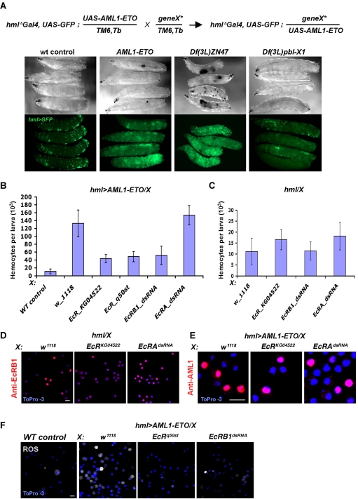
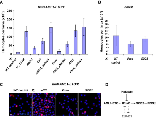
Among mutations in human Runx1/AML1 transcription factors, the t(8;21)(q22;q22) genomic translocation that creates an AML1-ETO fusion protein is implicated in etiology of the acute myeloid leukemia. To identify genes and components associated with this oncogene we used Drosophila as a genetic model. Expression of AML1-ETO caused an expansion of hematopoietic precursors in Drosophila, which expressed high levels of reactive oxygen species (ROS). Mutations in functional domains of the fusion protein suppress the proliferative phenotype. In a genetic screen, we found that inactivation of EcRB1 or activation of Foxo and superoxide dismutase-2 (SOD2) suppress the AML1-ETO-induced phenotype by reducing ROS expression in the precursor cells. Our studies indicate that ROS is a signaling factor promoting maintenance of normal as well as the aberrant myeloid precursors and suggests the importance of antioxidant enzymes and their regulators as targets for further study in the context of leukemia.


Figures

Figure 1
Figure 1
AML1-ETO induces generation of ROS+, Wg+ hemocyte precursors, and increased proliferation of hemocytes in larval circulation. (A) AML1-ETO, AML1, ETO, and Lz were expressed under control of the hemocyte specific driver hmlΔ-Gal4, using Gal4/UAS system. Hemocytes from 3rd instar larva were extracted and counted for each genotype. hmlΔ-Gal4,UAS-GFP heterozygous were used as wild-type control (WT). AML1-ETO causes a robust increase in the number of hemocytes in larval circulation compared with wild-type and overexpression of the other proteins. (B) Hemocyte precursors, identified as cells that do not express maturation markers: P1, Pxn, PPO, and L1 (all green) are rarely seen in wild-type (rare examples shown by arrow), but are significantly elevated upon AML1-ETO expression (arrows). (C) Quantitation of the data in panel B (n = 10, P < .001). (D) Wg is highly expressed in rare precursor cells that are hml negative (arrowhead) in circulation of wild-type larvae (WT). Number of Wg+ hemocytes is significantly increased in AML1-ETO mutant background. (E) Quantitation of the data in panel D (n = 10, P < .001). (F) ROS is expressed in hmllow(1) and rarely at low levels in some hml+(2) hemocytes in circulation of wild-type larva (WT). Number of cells expressing high level of ROS is increased in mutant background (AML1-ETO). (G) Quantitation of the data in panel F (n = 10, P < .001). (H) ROS+ cells (indicated by arrowhead) in WT and AML1-ETO backgrounds fail to phagocytose FluoSpheres (FS, blue) microparticles, suggesting they are precursors. Hemocyte markers, ROS, ToPro-3, and microspheres dyes color-coded in left panels. Scale bars, 5 μm.
Figure 2
Figure 2
Functional domains of AML1-ETO are critical for its activity in Drosophila blood cells. (A) Schematic representation of the AML1-ETO domains and mutations. Interaction sites of AML1-ETO with DNA, CBFβ (light blue lines), and HDAC1-3, Sin3A, N-CoR/SMRT, ETO, and SON (dark blue lines) are indicated below the protein scheme. (B) UAS constructs encoding wild-type AML1-ETO and its mutant variants (indicated on panel) were expressed under control of hmlΔ-Gal4. hmlΔ-Gal4,UAS-GFP heterozygous were used as wild-type control (WT control). In contrast to intact AML1-ETO, mutant forms with altered DNA-binding (R174Q), CBFβ-binding [Y113A/T161A (YT)], or both DNA- and CBFβ-binding [R174Q/Y113A/T161A (YTR)] domains failed to induce proliferation of hemocytes (n = 10, P < .001). Truncated AML1-ETO(NHR3X) and (NHR2X) proteins were less active in inducing hemocyte proliferation than intact protein (n = 10, P < .001). The AML1-ETO(NHR2X542,m7) mutant with disrupted HHR, NHR3, and MYND domains was unable to induce hemocyte proliferation.
Figure 3
Figure 3
A genetic screen identified ecdysone receptors as modifiers of AML1-ETO induced blood disorder. (A) Schema of modifier genetic screen: flies of synthesized genetic background containing hmlΔ-Gal4,UAS-GFP;UAS-AML1-ETO/TM6,Tb were crossed with mutant alleles. Larvae from these crosses were scored under direct (top panels) and fluorescent (bottom panels) illumination (Zeiss SteREO Lumar.V12 Stereomicroscope, 12× magnification); from left to right: normal density of GFP+ hemocytes in wild-type (WT) larvae of hmlΔ-Gal4,UAS-GFP background. Numbers of GFP+ hemocytes and black melanotic tumors are robustly elevated in hmlΔ-Gal4,UAS-GFP;UAS-AML1-ETO larvae (AML1-ETO). Examples of enhancer and suppressor modifiers of AML1-ETO phenotype: single copy deficiencies [Df(3L)ZN47] or [Df(3L)pbl-X1] cause a dramatic increase and decrease, respectively, in the number of circulating hemocytes and melanotic tumors in hmlΔ-Gal4, UAS-GFP; UAS-AML1-ETO larvae. These deficiencies include hundreds of genes, a majority of which remain yet uncharacterized. Analysis of available mutant alleles of genes belonging to these genomic intervals has not yet identified single loci that can modify the phenotype. (B) EcR-B1 is required for induction of hemocyte proliferation by AML1-ETO. EcR-B1 and EcR-A were inactivated in hemocytes of hmlΔ-Gal4,UAS-GFP;UAS-AML1-ETO background (hml > AML1-ETO). AML1-ETO induced proliferation of hemocytes was dramatically suppressed by EcR hemizygosity (alleles KG04522, q50st) and dsRNA of EcR-B1. In contrast,the oncogene induced proliferation of hemocytes was very mildly increased by dsRNA allele of EcR-A. hmlΔ-Gal4,UAS-GFP heterozygous were used as wild-type control (WT control). (C) As controls for (B), hemizygosity for EcR-B1 or inactivation of either EcR-B1 or EcR-A with corresponding dsRNAs in normal hemocytes [hmlΔ-Gal4,UAS-GFP(hml)] does not affect proliferation of otherwise hemocytes. (D) Inactivation of EcR-A causes increase in circulating hemocytes expressing EcR-B1. EcR-B1 is highly expressed in nuclei of a population of circulating hemocytes in wild-type larvae (hml/w1118). Expression of EcR-B1 is significantly reduced in EcR KG04522 heterozygous (hml/EcRKG04522) animals, while the number of cells expressing high level of EcR-B1 is significantly increased upon dsRNA-mediated inactivation of EcR-A (hml/EcR-AdsRNA). (E) Nuclear localization of AML1-ETO in hemocytes is not affected by deficiency of EcR-B1 (EcR KG04522) or EcR-A (EcR-AdsRNA) in AML1-ETO mutant background. (G) Number of ROS+ hemocyte precursors is significantly reduced upon inactivation of EcR-B1 in AML1-ETO mutant larvae (hml > AML1-ETO). Abbreviations of genotypes indicated on top of each panel, cell markers are color coded. Scale bars, 5 μm.
Figure 4
Figure 4
SOD2 activation by Foxo suppresses generation of AML1-ETO–induced precursors. (A) Activation of SOD2 or Catalase or Foxo, or inactivation of Akt1 causes suppression of AML1-ETO–mediated hemocyte proliferation. Corresponding UAS constructs (indicated on x-axis) were expressed in hemocytes expressing AML1-ETO (hmlΔ-Gal4,UAS-GFP;UAS-AML1-ETO: hml > AML1-ETO). Number of hemocytes in AML1-ETO mutant was significantly reduced by ectopic expression of Foxo, AktdsRNA, SOD2, or Catalase (n = 10, P < .001). Number of hemocytes in AML1-ETO mutant was not significantly affected by overexpression of Akt1, ptendsRNA, SOD2dsRNA. hmlΔ-Gal4,UAS-GFP heterozygous were used as wild-type control (WT control). (B) In a wild-type background, the number of hemocytes is not significantly affected by overexpression of SOD2 or Foxo with hmlΔ-Gal4,UAS-GFP. (C) Activation of SOD2 or Foxo causes significant reduction of Wg+ hemocyte precursors in AML1-ETO mutant. SOD2 or Foxo were expressed in hemocytes expressing AML1-ETO (hml > AML1-ETO). Scale bars, 5 μm. (D) A schematic diagram depicting the relationship between PI3K/Akt and EcR-B1 pathways in negative regulation of FoxO that is required for positive regulation of ROS-inactivating enzymes. We propose that AML1-ETO suppresses FoxO function, leading to an increase of ROS in hemocyte precursors.

References

    1. Canon J, Banerjee U. Runt and Lozenge function in Drosophila development. Semin Cell Dev Biol. 2000;11(5):327–336. - PubMed
    1. Ito Y. RUNX genes in development and cancer: regulation of viral gene expression and the discovery of RUNX family genes. Adv Cancer Res. 2008;99:33–76. - PubMed
    1. Kalev-Zylinska ML, Horsfield JA, Flores MV, et al. Runx1 is required for zebrafish blood and vessel development and expression of a human RUNX1-CBF2T1 transgene advances a model for studies of leukemogenesis. Development. 2002;129(8):2015–2030. - PubMed
    1. de Bruijn MF, Speck NA. Core-binding factors in hematopoiesis and immune function. Oncogene. 2004;23(24):4238–4248. - PubMed
    1. Watanabe-Okochi N, Kitaura J, Ono R, et al. AML1 mutations induced MDS and MDS/AML in a mouse BMT model. Blood. 2008;111(8):4297–4308. - PubMed

Publication types

MeSH terms

LinkOut - more resources