. 2010 Sep 17;329(5998):1526-30.
doi: 10.1126/science.1190187. Epub 2010 Aug 5.

Structural basis for activation of class Ib ribonucleotide reductase

Affiliations

Structural basis for activation of class Ib ribonucleotide reductase

Amie K Boal et al. Science. .

Abstract

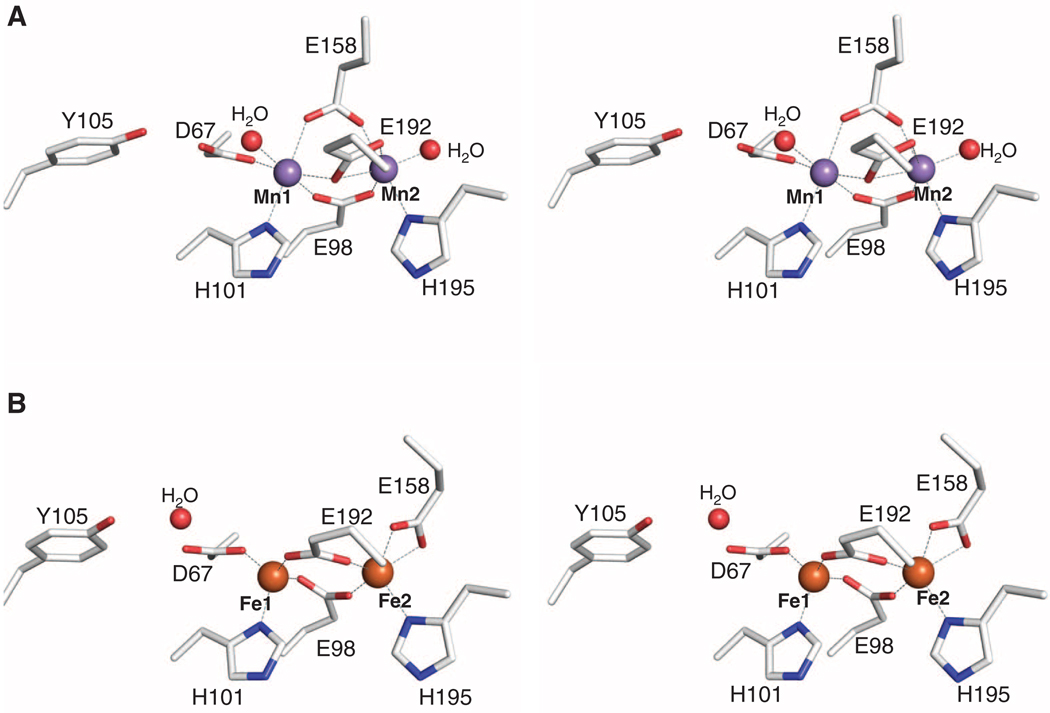
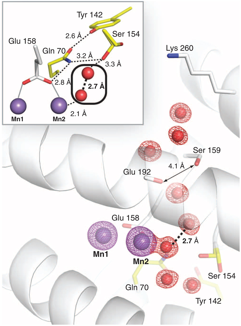
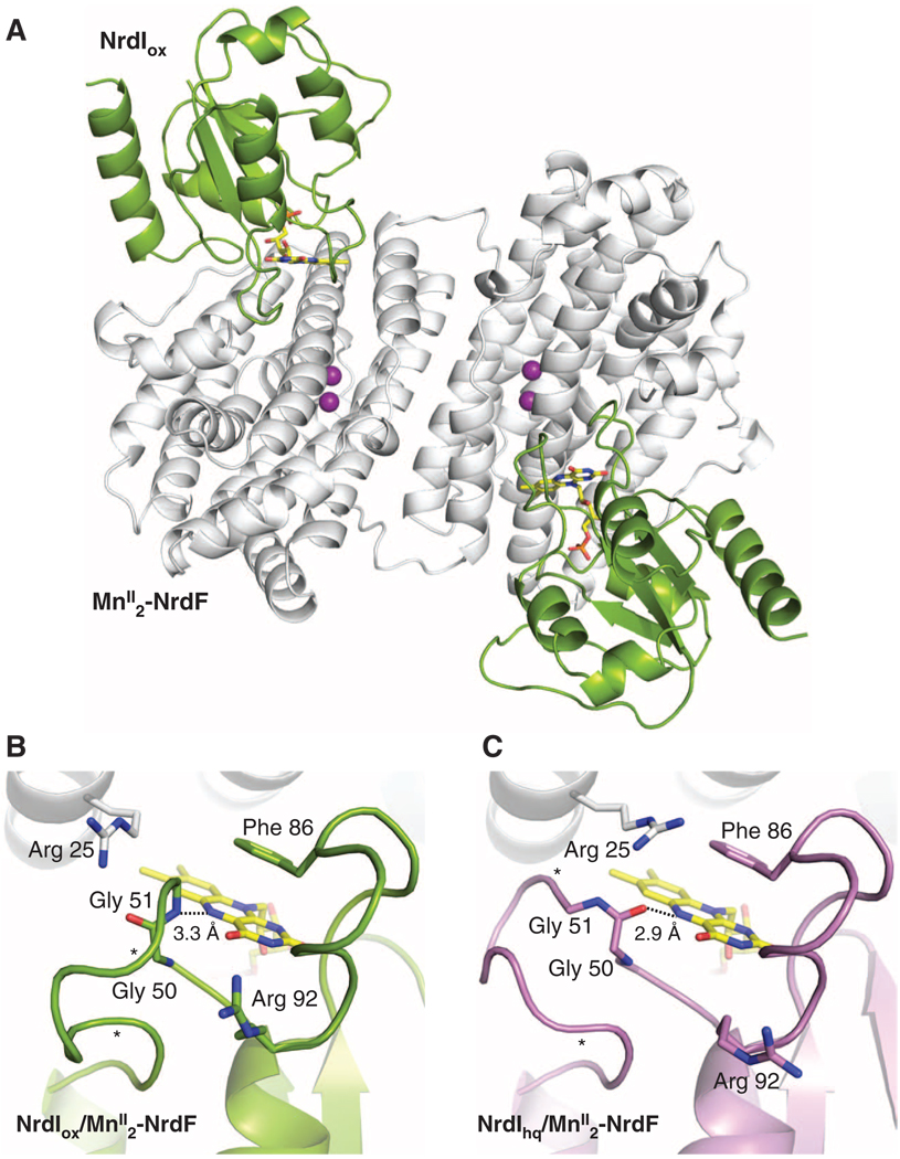
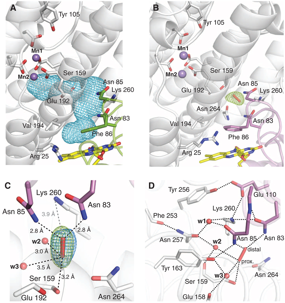
The class Ib ribonucleotide reductase of Escherichia coli can initiate reduction of nucleotides to deoxynucleotides with either a Mn(III)2-tyrosyl radical (Y•) or a Fe(III)2-Y• cofactor in the NrdF subunit. Whereas Fe(III)2-Y• can self-assemble from Fe(II)2-NrdF and O2, activation of Mn(II)2-NrdF requires a reduced flavoprotein, NrdI, proposed to form the oxidant for cofactor assembly by reduction of O2. The crystal structures reported here of E. coli Mn(II)2-NrdF and Fe(II)2-NrdF reveal different coordination environments, suggesting distinct initial binding sites for the oxidants during cofactor activation. In the structures of Mn(II)2-NrdF in complex with reduced and oxidized NrdI, a continuous channel connects the NrdI flavin cofactor to the NrdF Mn(II)2 active site. Crystallographic detection of a putative peroxide in this channel supports the proposed mechanism of Mn(III)2-Y• cofactor assembly.


Figures

Fig. 1
Fig. 1
Structures of MnII2-NrdF and FeII2-NrdF. (A) Stereoview of the MnII2-NrdF active site. MnII ions are shown as purple spheres, water molecules are shown as red spheres, and NrdF side chains are represented in stick format and colored by atom type. (B) Stereoview of the FeII2-NrdF active site. FeII ions, modeled at 0.5 occupancy, are shown as orange spheres. Metal-ligand interactions are highlighted with dashed lines.
Fig. 2
Fig. 2
The MnII2-NrdF solvent-exposed active-site channel terminating at Mn2. A 2FoFc electron density map (red mesh, contoured at 2σ) shows ordered waters in the channel. The Mn anomalous difference Fourier map (purple mesh, contoured at 12σ) is also shown. Residues implicated in channel access are shown as white sticks, and a conserved hydrogen bonding network (illustrated with dashed lines in inset) linking ordered solvent in the channel to Mn2 ligand Glu158 is shown as yellow sticks. Ser154 is modeled in two separate rotamer conformations in MnII2-NrdF, but in all NrdI-NrdF complex structures, it adopts the rotamer that points into the solvent channel.
Fig. 3
Fig. 3
Structures of NrdI-NrdF protein-protein complexes. (A) A ribbon diagram of the NrdIox-NrdF structure. NrdI is shown in green and MnII2-NrdF is shown in white. The NrdI FMN cofactor is shown as yellow sticks. (B) The NrdI FMN environment in the NrdIox-NrdF structure (NrdI shown in green). (C) The NrdI FMN environment in the NrdIhq-NrdF structure (NrdI shown in purple). Hydrogen bonding interactions with the FMN N5 position are shown as dashed lines. The electron density for the 50s loop is not completely continuous between the asterisks.
Fig. 4
Fig. 4
The NrdI-NrdF channel. (A) NrdI-NrdF complex formation extends the NrdF active-site channel to the FMN cofactor. The complex channel is depicted as a light blue mesh and was calculated using a 1.4 Å probe radius. Selected NrdI (green) and NrdF (white) residues lining the channel are shown as sticks. (B) Observation of a trapped species, best modeled as peroxide, in the NrdI-NrdF channel in a crystal reduced by dithionite in the presence of oxygen (NrdIhq-NrdFperox). Strong FoFc electron density (green mesh, contoured at 3.3σ) is present in the channel after the first refinement cycle. The FMN cofactor (yellow), NrdI side chains lining the channel (purple), NrdF residues in the channel and at the active site (white), and the peroxide (red) are all shown as sticks. (C) A magnified view of the modeled peroxide shown in Fig. 3B and hydrogen bonding interactions with residues and solvent in the channel. The final 2FoFc electron density (blue mesh, contoured at 1.8σ) is superimposed on the initial FoFc electron density map from Fig. 3A. Water molecules are shown as red spheres. Dashed black lines indicate potential hydrogen bonding interactions. The gray dashed line represents the distance between the modeled peroxide and the nearest charged residue, conserved NrdF residue Lys260. The Glu192 backbone carbonyl group and the side chain of Ser159 constitute the narrowest point of the active-site channel. The oxygen atom distal to the MnII2 site interacts strongly with the side chains of NrdI residues Asn85 (2.8 Å) and conserved Asn83 (2.8 Å). (D) The extended hydrogen bonding network near the putative peroxide binding site. The side-chain orientations of Asn83 and Asn257 can be assigned unequivocally based on their interactions with Lys260 and the backbone amide nitrogen of Asn85, and the carbonyl oxygen of Phe253, respectively. The interactions of w2 with Asn257 (2.8 Å) and the backbone carbonyl of Ser159 (2.7 Å) constrain w2 to act as a hydrogen bond acceptor for the proximal oxygen atom of the modeled peroxide (2.9 Å), suggesting that this oxygen is protonated. The distal oxygen accepts two hydrogen bonds from Asn83 and Asn85. Because no other potential hydrogen bond donor or acceptor exists for this oxygen atom, its protonation state cannot be determined from this analysis.

Comment in

  • Biochemistry. A never-ending story.
    Sjöberg BM. Sjöberg BM. Science. 2010 Sep 17;329(5998):1475-6. doi: 10.1126/science.1196347. Science. 2010. PMID: 20847256 No abstract available.

Similar articles

Cited by

References

    1. Nordlund P, Reichard P. Annu. Rev. Biochem. 2006;75:681. - PubMed
    1. Gon S, Faulkner MJ, Beckwith J. Antioxid. Redox Signal. 2006;8:735. - PubMed
    1. McHugh JP, et al. J. Biol. Chem. 2003;278:29478. - PubMed
    1. Monje-Casas F, Jurado J, Prieto-Alamo MJ, Holmgren A, Pueyo C. J. Biol. Chem. 2001;276:18031. - PubMed
    1. Atkin CL, Thelander L, Reichard P, Lang G. J. Biol. Chem. 1973;248:7464. - PubMed

Publication types

MeSH terms

LinkOut - more resources