. 2010 Oct;299(4):G887-97.
doi: 10.1152/ajpgi.00216.2010. Epub 2010 Aug 5.

Identification of microRNAs during rat liver regeneration after partial hepatectomy and modulation by ursodeoxycholic acid

Affiliations

Identification of microRNAs during rat liver regeneration after partial hepatectomy and modulation by ursodeoxycholic acid

Rui E Castro et al. Am J Physiol Gastrointest Liver Physiol. 2010 Oct.

Abstract

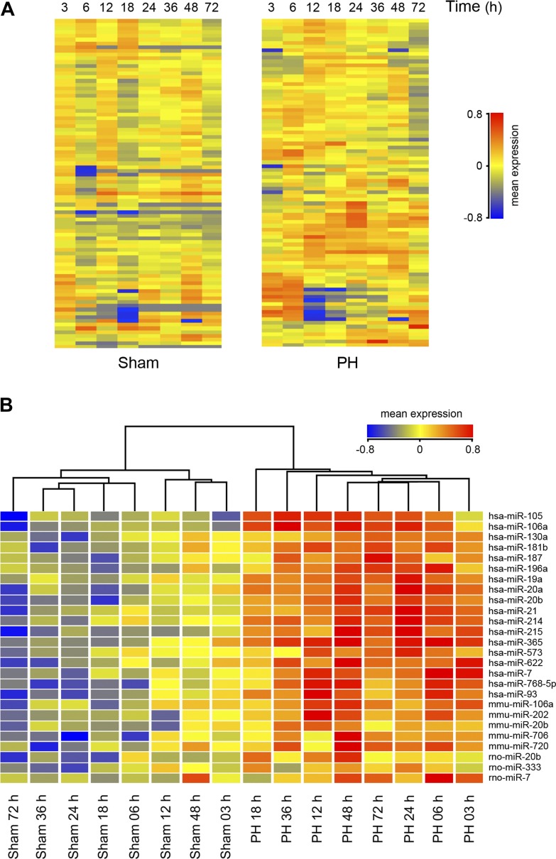
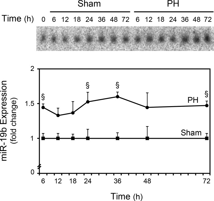
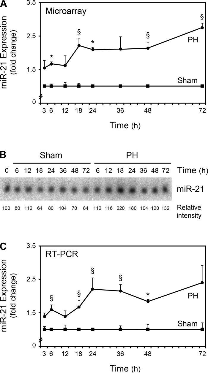
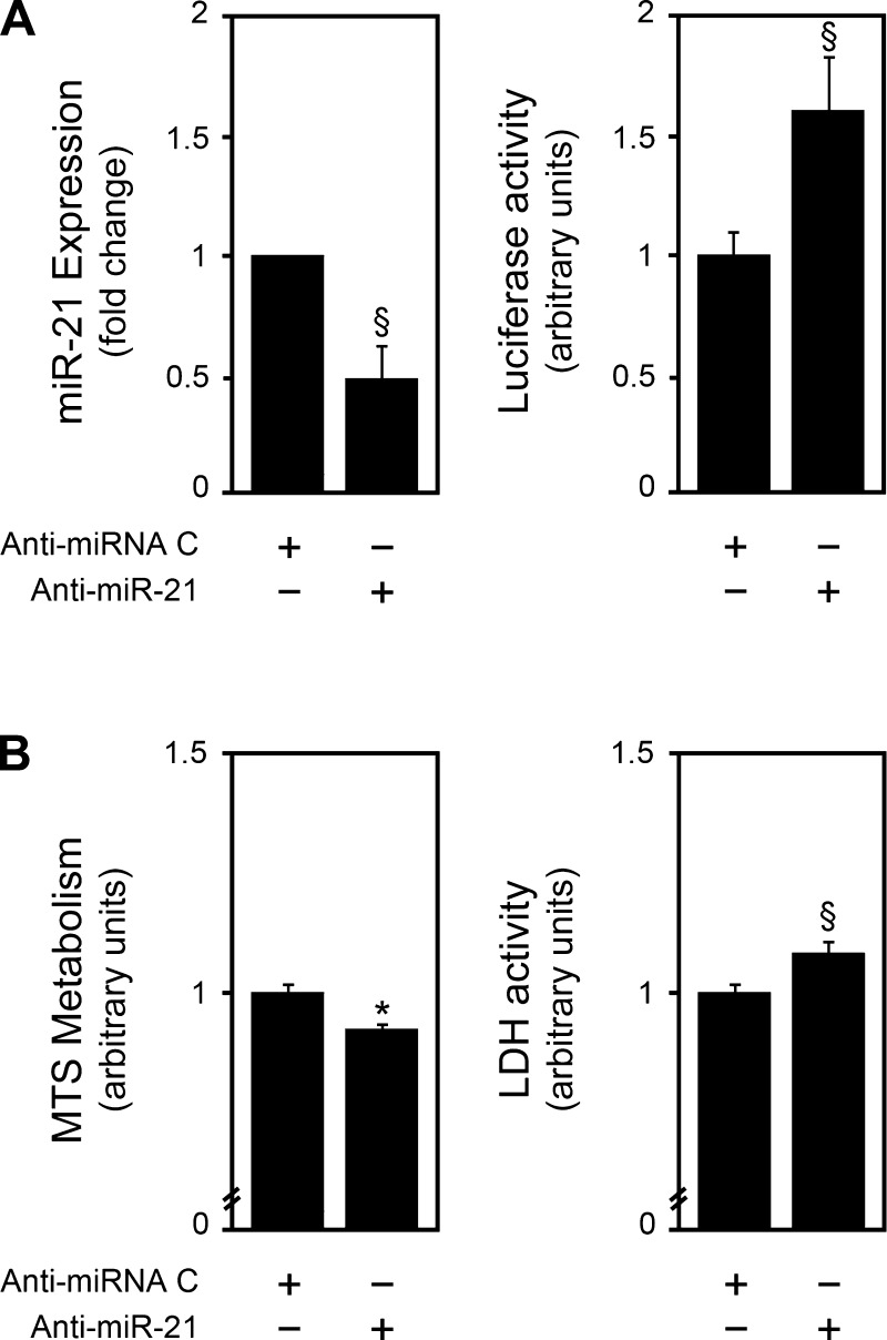
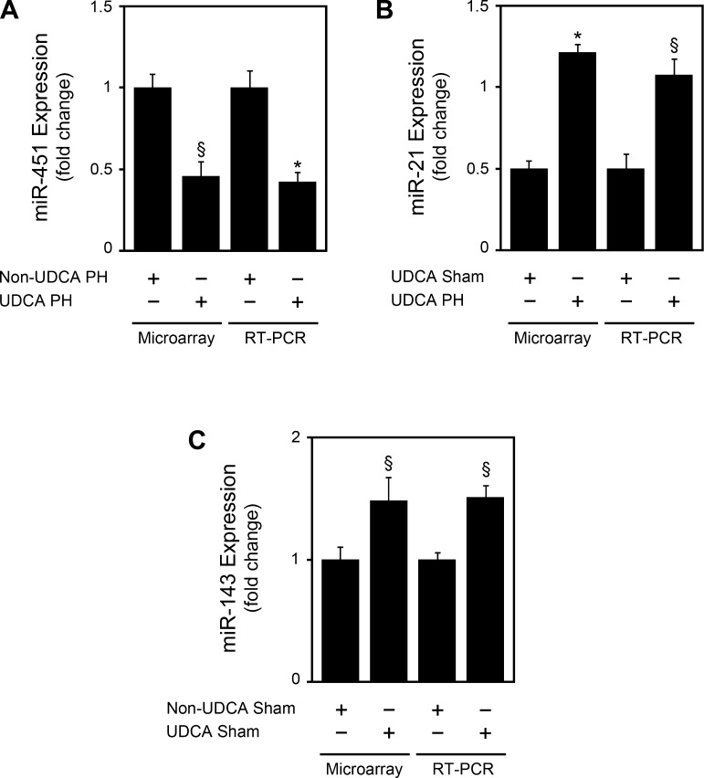
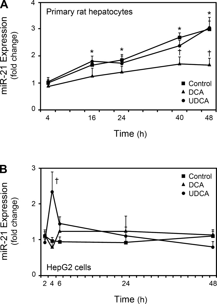
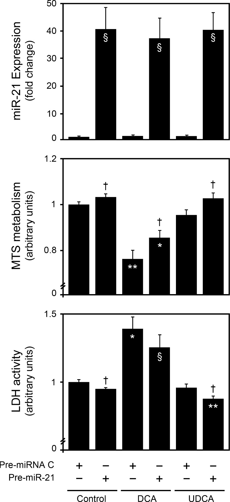
New gene regulation study tools such as microRNA (miRNA or miR) analysis may provide unique insights into the remarkable ability of the liver to regenerate. In addition, we have previously shown that ursodeoxycholic acid (UDCA) modulates mRNA levels during liver regeneration. Bile acids are also homeotrophic sensors of functional hepatic capacity. The present study was designed to determine whether miRNAs are modulated in rats following 70% partial hepatectomy (PH) and elucidate the role of UDCA in regulating miRNA expression during liver regeneration (LR). Total RNA was isolated from livers harvested at 3-72 h following 70% PH or sham operations, from both 0.4% (wt/wt) UDCA and control diet-fed animals. By using a custom microarray platform we found that several miRNAs are significantly altered after PH by >1.5-fold, including some previously described as modulators of cell proliferation, differentiation, and death. In particular, expression of miR-21 was increased after PH. Functional modulation of miR-21 in primary rat hepatocytes increased cell proliferation and viability. Importantly, UDCA was a strong inducer of miR-21 both during LR and in cultured HepG2 cells. In fact, UDCA feeding appeared to induce a sustained increase of proliferative miRNAs observed at early time points after PH. In conclusion, miRNAs, in particular miR-21, may play a significant role in modulating proliferation and cell cycle progression genes after PH. miR-21 is additionally induced by UDCA in both regenerating rat liver and in vitro, which may represent a new mechanism behind UDCA biological functions.


Figures

Fig. 1.
Fig. 1.
Differentially expressed liver microRNAs (miRNAs or miRs) in partial hepatectomy (PH)- vs. sham-operated rats, from 3 to 72 h after PH. A: distribution of 88 distinct miRNAs in the regenerating liver. These miRNA probes represent the transcripts that gave the strongest normalized hybridization signals in all sampled populations. Overall, miRNA expression was increased in PH samples. B: hierarchical clustering of miRNA genes whose expression was significantly altered by >1.5-fold in at least 1 time point after PH. Each row represents an individual gene and each column represents 3 RNA liver samples from 3 different animals. Results are presented to highlight the clustering analysis, with the most distant groups on the left and right extremes of the hierarchical tree. The relative abundance of individual miRNAs in different time point populations is presented as heat maps, with the lowest and highest intensity values in blue and red, respectively. The scale is shown at right, with the relative abundance in arbitrary units.
Fig. 2.
Fig. 2.
Northern blot analysis of miRNA-19b. Northern blots were performed as described in materials and methods. Blots were normalized to 5S rRNA and results are expressed as mean ± SE fold change for 3 different animals. A representative Northern blot is shown. §P < 0.05 from the corresponding controls.
Fig. 3.
Fig. 3.
miR-21 expression is increased after PH. Expression of miR-21 from 3 to 72 h after PH was evaluated by microarray (A), Northern blot (B), and real-time RT-PCR (C) analyses as described in materials and methods. A representative Northern blot is shown. Results are expressed as mean ± SE fold change for 3 different animals. *P < 0.01 and §P < 0.05 from the corresponding controls.
Fig. 4.
Fig. 4.
miR-21 modulates cell proliferation and viability in primary rat hepatocytes. Primary rat hepatocytes were transfected with a miR-21 inhibitor as described in materials and methods. miR-21 silencing (A) was assessed by real-time RT-PCR (left) or luciferase activity (right) assays. Cell proliferation and viability (B) were assessed by the 3-(4,5-dimethylthiazol-2-yl)-5-(3-carboxymethoxyphenyl)-2-(4-sulfophenyl)-2H-tetrazolium (MTS) metabolism (left) and lactate dehydrogenase (LDH) activity (right) assays, respectively. Results are expressed as means ± SE from at least 4 different experiments. §P < 0.05 and *P < 0.01 from the corresponding controls.
Fig. 5.
Fig. 5.
Ursodeoxycholic acid (UDCA) modulates miR-451, -21, and -143 expression in accordance with microarray results. miRNA expression was evaluated by real-time RT-PCR as described in materials and methods. A: miR-451 expression in UDCA diet-fed animals at 36 h after PH, compared with control diet PH animals. B: miR-21 expression in UDCA-fed animals at 36 h after PH, compared with sham animals. C: miR-143 expression in sham UDCA-diet-fed animals, at 36 h after PH, compared with control diet sham animals. Results are expressed as means ± SE of 3 different animals. *P < 0.01 and §P < 0.05 from the corresponding controls.
Fig. 6.
Fig. 6.
UDCA and deoxycholic acid (DCA) modulate miR-21 expression in proliferating HepG2 cells and primary rat hepatocytes, respectively. Cells were incubated with 100 μM UDCA, or DCA, or no addition (control) for 4, 16, 24, 40, and 48 h in primary rat hepatocytes (A), or 2, 4, 6, 24, and 48 h in HepG2 cells (B). Total RNA was obtained for real-time RT-PCR analysis as described in materials and methods. Results are expressed as means ± SE for at least 3 different experiments. *P < 0.01 from control 4 h and †P < 0.05 from the respective control.
Fig. 7.
Fig. 7.
Bile acids modulate cell proliferation and viability in primary rat hepatocytes, in part, through miR-21. Primary rat hepatocytes were transfected with a miR-21 precursor and incubated with 100 μM UDCA, or DCA, or no addition (control) as indicated in materials and methods. miR-21 overexpression was assessed by real-time RT-PCR (top). Cell proliferation and viability were assessed by the MTS metabolism (middle) and LDH activity (bottom) assays, respectively. Results are expressed as means ± SE from at least 3 different experiments. §P < 0.05, *P < 0.01 and **P < 0.001 from the no-bile-acid pre-miRNA control. †P < 0.05 from the corresponding pre-miRNA controls.

References

    1. Amaral JD, Castro RE, Solá S, Steer CJ, Rodrigues CM. p53 is a key molecular target of ursodeoxycholic acid in regulating apoptosis. J Biol Chem 282: 34250–34259, 2007. - PubMed
    1. Bandres E, Bitarte N, Arias F, Agorreta J, Fortes P, Agirre X, Zarate R, Diaz-Gonzalez JA, Ramirez N, Sola JJ, Jimenez P, Rodriguez J, Garcia-Foncillas J. microRNA-451 regulates macrophage migration inhibitory factor production and proliferation of gastrointestinal cancer cells. Clin Cancer Res 15: 2281–2290, 2009. - PubMed
    1. Barone M, Francavilla A, Polimeno L, Ierardi E, Romanelli D, Berloco P, Di Leo A, Panella C. Modulation of rat hepatocyte proliferation by bile salts: in vitro and in vivo studies. Hepatology 23: 1159–1166, 1996. - PubMed
    1. Bockhorn M, Goralski M, Prokofiev D, Dammann P, Grunewald P, Trippler M, Biglarnia A, Kamler M, Niehues EM, Frilling A, Broelsch CE, Schlaak JF. VEGF is important for early liver regeneration after partial hepatectomy. J Surg Res 138: 291–299, 2007. - PubMed
    1. Bostjancic E, Glavac D. Importance of microRNAs in skin morphogenesis and diseases. Acta Dermatovenerol Alp Panonica Adriat 17: 95–102, 2008. - PubMed

Publication types

LinkOut - more resources