. 2010 Oct 1;107(7):851-9.
doi: 10.1161/CIRCRESAHA.109.215269. Epub 2010 Aug 5.

Cardiac small conductance Ca2+-activated K+ channel subunits form heteromultimers via the coiled-coil domains in the C termini of the channels

Affiliations

Cardiac small conductance Ca2+-activated K+ channel subunits form heteromultimers via the coiled-coil domains in the C termini of the channels

Dipika Tuteja et al. Circ Res. .

Abstract

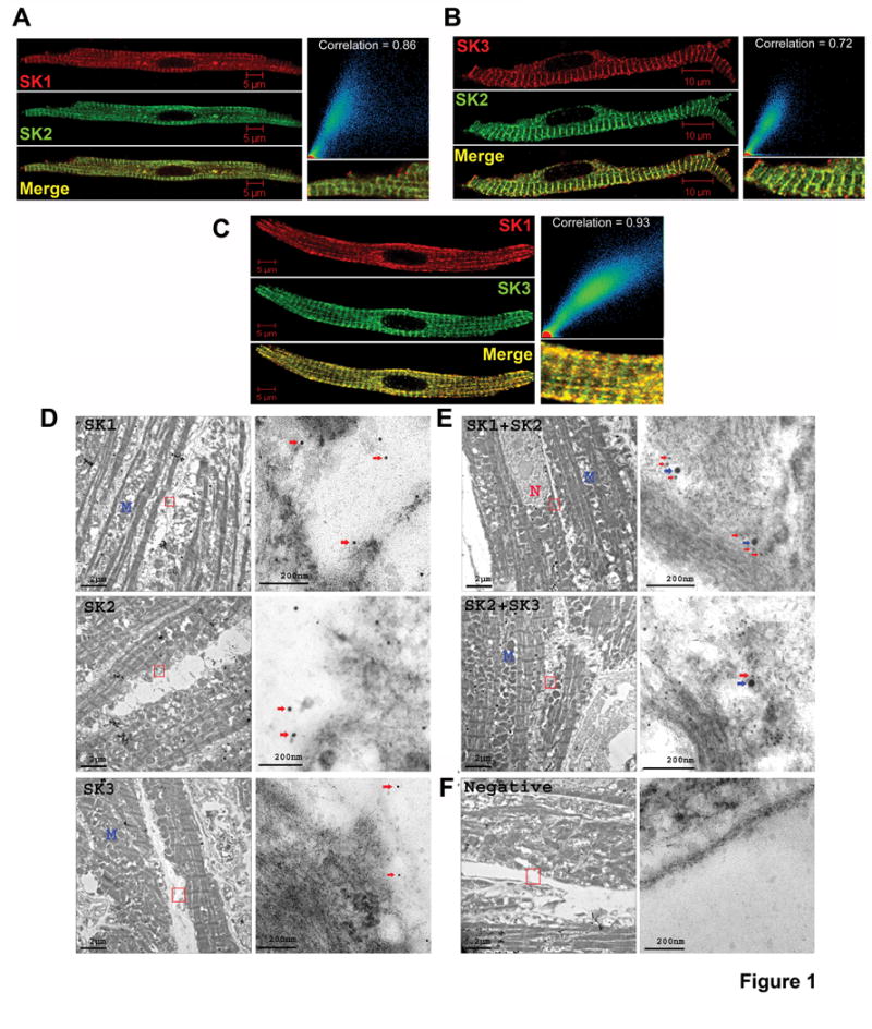
Rationale: Ca(2+)-activated K(+) channels are present in a wide variety of cells. We have previously reported the presence of small conductance Ca(2+)-activated K(+) (SK or K(Ca)) channels in human and mouse cardiac myocytes that contribute functionally toward the shape and duration of cardiac action potentials. Three isoforms of SK channel subunits (SK1, SK2, and SK3) are found to be expressed. Moreover, there is differential expression with more abundant SK channels in the atria and pacemaking tissues compared with the ventricles. SK channels are proposed to be assembled as tetramers similar to other K(+) channels, but the molecular determinants driving their subunit interaction and assembly are not defined in cardiac tissues.

Objective: To investigate the heteromultimeric formation and the domain necessary for the assembly of 3 SK channel subunits (SK1, SK2, and SK3) into complexes in human and mouse hearts.

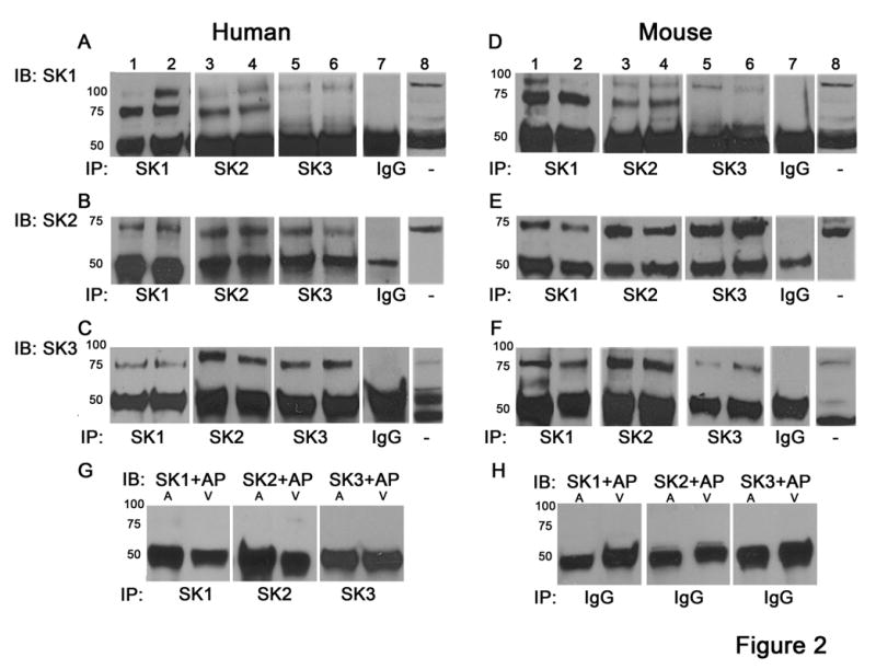
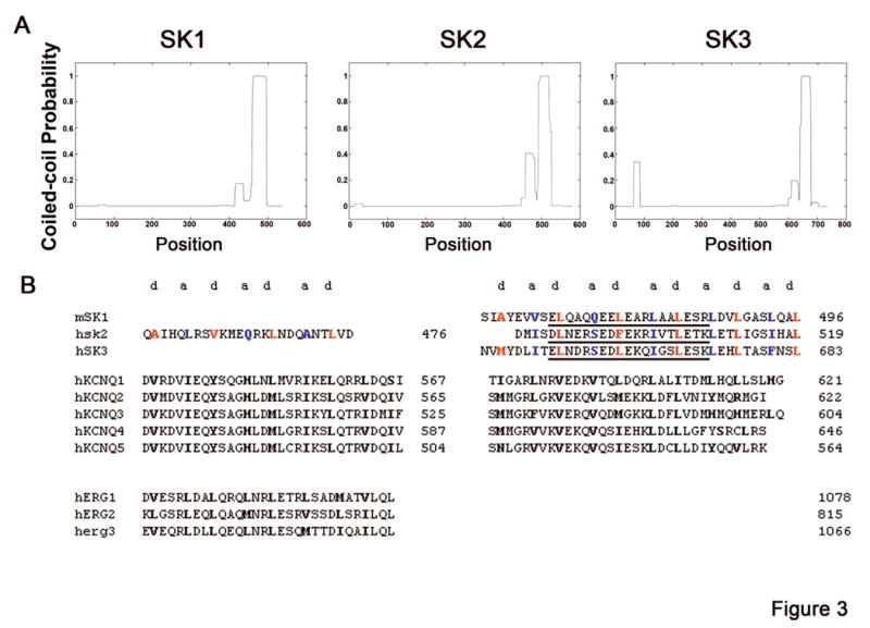
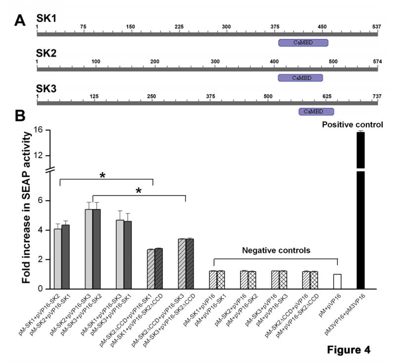
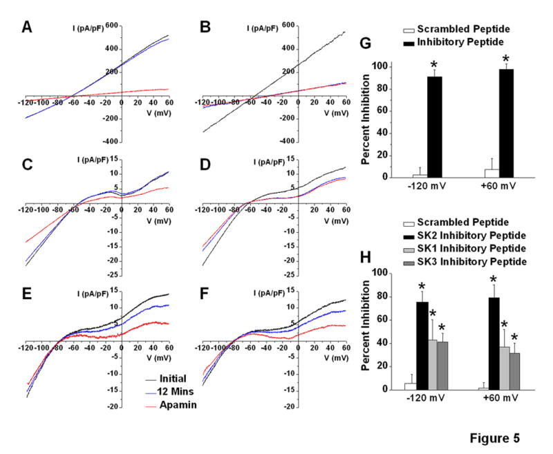
Methods and results: Here, we provide evidence to support the formation of heteromultimeric complexes among different SK channel subunits in native cardiac tissues. SK1, SK2, and SK3 subunits contain coiled-coil domains (CCDs) in the C termini. In vitro interaction assay supports the direct interaction between CCDs of the channel subunits. Moreover, specific inhibitory peptides derived from CCDs block the Ca(2+)-activated K(+) current in atrial myocytes, which is important for cardiac repolarization.

Conclusions: The data provide evidence for the formation of heteromultimeric complexes among different SK channel subunits in atrial myocytes. Because SK channels are predominantly expressed in atrial myocytes, specific ligands of the different isoforms of SK channel subunits may offer a unique therapeutic opportunity to directly modify atrial cells without interfering with ventricular myocytes.


Figures

Figure 1
Figure 1. Subcellular distribution of SK1, SK2, and SK3 channel subunits in isolated mouse atrial myocytes using immunohistochemistry and immune-EM
Confocal photomicrographs of single isolated mouse atrial myocytes doubly stained with anti-SK antibodies in different combinations as follows: (A) goat anti-SK1 and rabbit anti-SK2, (B) rabbit anti-SK2 and goat anti-SK3, and (C) rabbit anti-SK1 and mouse anti-SK3 antibodies. Scale bars are 5 μm except in panel B (10 μm). Merged images are shown in third row of each panel. Scatter plots in the right panels show high correlation between different SK channel subunits. All pixels in the images have been assigned a position on the Scatter plots and are placed according to the intensity of red or green color. In addition, merge images are shown at higher magnification in the right panels. D-E, Electron photomicrographs showing the ultra-structure of mouse atrial myocytes and subcellular distribution of SK1, SK2 and SK2 channel subunits. (D) Single immunolabeling for SK1, SK2 and SK3 channel subunits. Left, images taken under 3800× magnification with scale bars of 2 μm; Right, higher magnification images of areas marked by red boxes in the left panels with scale bars of 200 nm. Arrows indicate gold particles which are 10 nm, 15 nm and 5 nm for SK1, SK2 and SK3 staining, respectively. (E) Double labeling for SK1 and SK2, as well as SK2 and SK3 channel subunits. For SK1 and SK2 double labeling, 10 nm and 20 nm gold particle-conjugated secondary antibodies were used to localize SK1 and SK2 subunits, respectively. Note the clustering of SK1 (red arrows) and SK2 (blue arrows) channel subunits as shown by arrows. For SK2 and SK3, 20 nm and 10 nm gold-conjugated secondary antibodies were used to localize SK2 (blue arrows) and SK3 (red arrows) subunits, respectively. Note SK2 and SK3 channels are closely located as shown by arrows. (F) Negative controls are labeled by gold-conjugated secondary antibodies only and are devoid of any gold particles. N, nucleus; M, mitochondria.
Figure 2
Figure 2. Heteromultimeric protein complexes of SK1, SK2, and SK3 channels detected from atrial and ventricular tissues using co-IP
Human (A-C) and mouse (D-F) atrial (Lanes 1, 3, 5) and ventricular (Lanes 2, 4, 6) tissue homogenates were immunoprecipitated (IP) with anti-SK antibodies, proteins were eluted and Western blot analysis (IB) was performed. The experiments were repeated a total of 6 times. (A,D) Immunoprecipitation of SK1 channel protein by anti-SK2 as well as anti-SK3 antibodies coupled to agarose beads, represented by a 70 kD band in the immunoblot. Both monomers and dimers were detected as distinct bands. (B,E) Immunoprecipitation of SK2 channel protein by anti-SK1 as well as anti-SK3 antibodies coupled to agarose beads, represented by a 70 kD band in the immunoblot. (C,F) Immunoprecipitation of SK3 channel protein by anti-SK2 as well as anti-SK3 antibodies coupled to agarose beads, represented by >70 kD band in the immunoblot. Negative control was performed by immunoblotting of eluted proteins immunoprecipitated with non-specific IgG (Lane 7, A-F) while positive control was performed using tissue homogenates (Lane 8, A-F). Lane 7 & 8 contained mixture of atrial and ventricular homogenates. (G) Control experiments were performed using mouse atrial (A) and ventricular (V) tissue homogenates immunoprecipitated (IP) with anti-SK antibodies, proteins were eluted and Western blot analysis (IB) was performed using anti-SK antibodies pre-incubated with their corresponding antigenic peptide (AP) in a ratio of 1:5. (H). Additional control experiments were performed using mouse atrial (A) and ventricular (V) tissue homogenates immunoprecipitated (IP) with non-specific normal rabbit anti-IgG antibodies, proteins were eluted and Western blot analysis (IB) was performed with anti-SK antibodies. In both G & H, except for heavy chain band of 50 kD, no other bands were detected.
Figure 3
Figure 3. Tetramerizing Coiled-coil domains (CCDs) in SK1, 2 and 3 channels
(A) Coiled-coil probability for cardiac SK channel proteins using COILS with scanning window of 21. (B) Amino acid sequence alignment of C-termini from mammalian SK1, SK2, SK3, KCNQ (Kv7) and hERG (Kv11.1) channels demonstrating the presence of bimodular TCC domains. “a” and “d” coiled-coil positions are indicated and color coded as blue and red, respectively in second CCD of SK2 channel protein. Underlined amino acid residues in SK1, SK2, and SK3 sequence denote the synthesized inhibitory peptides used in electrophysiological studies. m: mouse, h: human.
Figure 4
Figure 4. Identification of interaction between different SK channel subunits usingin vitrointeraction assay
Protein-protein interactions were assayed using the c-terminal fusion constructs of SK1-3 in pM and pVP16 vectors. (A) Schematic diagrams of cardiac SK1-SK3 proteins depicting the location of CaMBD. CaMBD and the region toward the C-terminus were used for the construction of the fusion proteins. (B) Protein-protein interactions among SK1, 2 and 3 C-termini are depicted by the fold increase of SEAP activity over controls. The roles of CCDs in SK2 channel in the interaction between different SK channel subunits were directly tested using SK2 C terminal constructs with deletion of the CCD domains (pM-SK2-ΔCCD and pVP16-SK2-ΔCCD).
Figure 5
Figure 5. Peptides derived from the SK1, SK2 and SK3 CCD inhibitIK,Cain atrial myocytes
A voltage-ramp protocol was applied from -120 to +60 mV at a holding potential of -55 mV. IK,Ca was recorded immediately after establishment of whole-cell mode (black line), 12 minutes thereafter (blue line) and after application of apamin (100 pM, red line). (A&B) IK,Ca density (pA/pF) recorded from tsA201 cells expressing SK2 current using scrambled (A) compared to SK2 (B) inhibitory peptides, respectively. (C-D) IK,Ca density recorded from mouse atrial myocytes using scrambled (C) compared to SK2 (D), SK1 (E), or SK3 (F) inhibitory peptides, respectively. (G&H) Summary data of percent inhibition of apamin-sensitive IK,Ca at -120 and +60 mV from tsA201 cells (G) and atrial myocytes (H), respectively (n=4-6), *p<0.05 comparing the inhibitory peptides to scrambled peptide.

References

    1. Kohler M, Hirschberg B, Bond CT, Kinzie JM, Marrion NV, Maylie J, Adelman JP. Small-conductance, calcium-activated potassium channels from mammalian brain. Science. 1996;273:1709–1714. - PubMed
    1. Stocker M. Ca2+-activated K+ channels: molecular determinants and function of the SK family. Nat Rev Neurosci. 2004;5:758–770. - PubMed
    1. Pedarzani P, McCutcheon JE, Rogge G, Jensen BS, Christophersen P, Hougaard C, Strobaek D, Stocker M. Specific enhancement of SK channel activity selectively potentiates the afterhyperpolarizing current IAHP and modulates the firing properties of hippocampal pyramidal neurons. J Biol Chem. 2005;280:41404–41411. - PubMed
    1. Xu Y, Tuteja D, Zhang Z, Xu D, Zhang Y, Rodriguez J, Nie L, Tuxson HR, Young JN, Glatter KA, Vazquez AE, Yamoah EN, Chiamvimonvat N. Molecular identification and functional roles of a Ca2+-activated K+ channel in human and mouse hearts. J Biol Chem. 2003;278:49085–49094. - PubMed
    1. Tuteja D, Xu D, Timofeyev V, Lu L, Sharma D, Zhang Z, Xu Y, Nie L, Vazquez AE, Young JN, Glatter KA, Chiamvimonvat N. Differential expression of small-conductance Ca2+-activated K+ channels SK1, SK2, and SK3 in mouse atrial and ventricular myocytes. Am J Physiol Heart Circ Physiol. 2005;289:H2714–2723. - PubMed

Publication types

MeSH terms