The relationship between cardiac output and dynamic cerebral autoregulation in humans
- PMID: 20689094
- PMCID: PMC2980368
- DOI: 10.1152/japplphysiol.01262.2009
The relationship between cardiac output and dynamic cerebral autoregulation in humans
Abstract
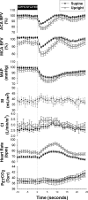
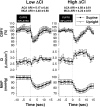
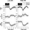
Cerebral autoregulation adjusts cerebrovascular resistance in the face of changing perfusion pressures to maintain relatively constant flow. Results from several studies suggest that cardiac output may also play a role. We tested the hypothesis that cerebral blood flow would autoregulate independent of changes in cardiac output. Transient systemic hypotension was induced by thigh-cuff deflation in 19 healthy volunteers (7 women) in both supine and seated positions. Mean arterial pressure (Finapres), cerebral blood flow (transcranial Doppler) in the anterior (ACA) and middle cerebral artery (MCA), beat-by-beat cardiac output (echocardiography), and end-tidal Pco(2) were measured. Autoregulation was assessed using the autoregulatory index (ARI) defined by Tiecks et al. (Tiecks FP, Lam AM, Aaslid R, Newell DW. Stroke 26: 1014-1019, 1995). Cerebral autoregulation was better in the supine position in both the ACA [supine ARI: 5.0 ± 0.21 (mean ± SE), seated ARI: 3.9 ± 0.4, P = 0.01] and MCA (supine ARI: 5.0 ± 0.2, seated ARI: 3.8 ± 0.3, P = 0.004). In contrast, cardiac output responses were not different between positions and did not correlate with cerebral blood flow ARIs. In addition, women had better autoregulation in the ACA (P = 0.046), but not the MCA, despite having the same cardiac output response. These data demonstrate cardiac output does not appear to affect the dynamic cerebral autoregulatory response to sudden hypotension in healthy controls, regardless of posture. These results also highlight the importance of considering sex when studying cerebral autoregulation.
Figures


References
-
- Aaslid R, Lindegaard KF, Sorteberg W, Nornes H. Cerebral autoregulation dynamics in humans. Stroke 20: 45–52, 1989 - PubMed
-
- Aaslid R, Markwalder TM, Nornes H. Noninvasive transcranial Doppler ultrasound recording of flow velocity in basal cerebral arteries. J Neurosurg 57: 769–774, 1982 - PubMed
-
- Bouma GJ, Muizelaar JP. Relationship between cardiac output and cerebral blood flow in patients with intact and with impaired autoregulation. J Neurosurg 73: 368–374, 1990 - PubMed
-
- Brown CM, Dutsch M, Hecht MJ, Neundorfer B, Hilz MJ. Assessment of cerebrovascular and cardiovascular responses to lower body negative pressure as a test of cerebral autoregulation. J Neurol Sci 208: 71–78, 2003 - PubMed
-
- Christie J, Sheldahl LM, Tristani FE, Sagar KB, Ptacin MJ, Wann S. Determination of stroke volume and cardiac output during exercise: comparison of two-dimensional and Doppler echocardiography, Fick oximetry, and thermodilution. Circulation 76: 539–547, 1987 - PubMed
Publication types
MeSH terms
LinkOut - more resources
Full Text Sources
Medical

