

Hemi-central retinal artery occlusion in young adults
- PMID: 20689202
- PMCID: PMC2992922
- DOI: 10.4103/0301-4738.67069
Hemi-central retinal artery occlusion in young adults
Abstract
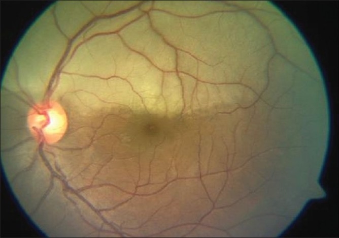
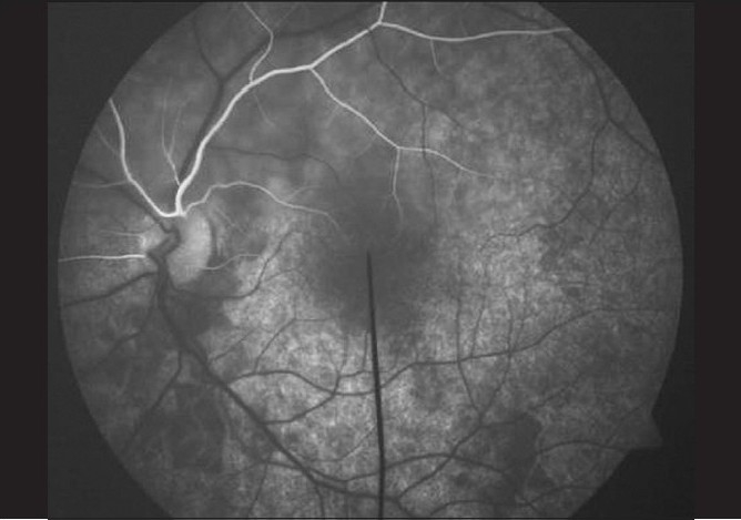
Amongst the clinical presentations of retinal artery occlusion, hemi-central retinal artery occlusion (Hemi-CRAO) is rarely described. This case series of four adults aged between 22 and 36 years attempts to describe the clinical profile, etiology and management of Hemi-CRAO. Case 1 had an artificial mitral valve implant. Polycythemia and malignant hypertension were noted in Case 2. The third patient had Leiden mutation while the fourth patient had Eisenmenger's syndrome. Clinical examination and fundus fluorescein angiography revealed a bifurcated central retinal artery at emergence from the optic nerve head, in all cases. Color Doppler examination of the central retinal artery confirmed branching of the artery behind the lamina cribrosa. It is hypothesized that bifurcation of central retinal artery behind the lamina cribrosa may predispose these hemi-trunks to develop an acute occlusion if associated with underlying risk factors. The prognosis depends upon arterial recanalisation and etiology of the thromboembolic event.
Figures


Comment in
-
Hemi-central retinal artery occlusion in young adults.Indian J Ophthalmol. 2011 May-Jun;59(3):261-2. doi: 10.4103/0301-4738.81033. Indian J Ophthalmol. 2011. PMID: 21586862 Free PMC article. No abstract available.
-
Hemicentral retinal artery occlusion in young adults.Indian J Ophthalmol. 2013 Jul;61(7):365. doi: 10.4103/0301-4738.97559. Indian J Ophthalmol. 2013. PMID: 23552353 Free PMC article. No abstract available.
Similar articles
-
Fundus changes in central retinal artery occlusion.Retina. 2007 Mar;27(3):276-89. doi: 10.1097/01.iae.0000238095.97104.9b. Retina. 2007. PMID: 17460582
-
Abnormal retinal vessel filling in central retinal artery occlusion.Clin Exp Optom. 2024 Nov;107(8):801-805. doi: 10.1080/08164622.2023.2298781. Epub 2024 Jan 7. Clin Exp Optom. 2024. PMID: 38184849
-
Central retinal artery occlusion.Indian J Ophthalmol. 2018 Dec;66(12):1684-1694. doi: 10.4103/ijo.IJO_1446_18. Indian J Ophthalmol. 2018. PMID: 30451166 Free PMC article. Review.
-
Combined central retinal vein and branch retinal artery occlusion in hyperhomocysteinaemia.BMJ Case Rep. 2016 Dec 14;2016:bcr2016218379. doi: 10.1136/bcr-2016-218379. BMJ Case Rep. 2016. PMID: 27974343 Free PMC article. No abstract available.
-
STRANGULATION-INDUCED CENTRAL RETINAL ARTERY OCCLUSION: CASE REPORT AND REVIEW OF THE LITERATURE.Retin Cases Brief Rep. 2017 Summer;11(3):258-260. doi: 10.1097/ICB.0000000000000334. Retin Cases Brief Rep. 2017. PMID: 27337704 Review.
Cited by
-
Central retinal artery occlusion with cilioretinal sparing in a patient with Eisenmenger syndrome.BMJ Case Rep. 2022 Feb 28;15(2):e246293. doi: 10.1136/bcr-2021-246293. BMJ Case Rep. 2022. PMID: 35228223 Free PMC article.
-
Hemicentral retinal artery occlusion in young adults.Indian J Ophthalmol. 2013 Jul;61(7):365. doi: 10.4103/0301-4738.97559. Indian J Ophthalmol. 2013. PMID: 23552353 Free PMC article. No abstract available.
-
A Case of Advanced Glaucoma with Increased Episcleral Venous Pressure in a 17-Year-Old with Eisenmenger Syndrome.Case Rep Ophthalmol Med. 2017;2017:5808047. doi: 10.1155/2017/5808047. Epub 2017 Oct 23. Case Rep Ophthalmol Med. 2017. PMID: 29201477 Free PMC article.
-
Hemi-central retinal artery occlusion following methanol toxicity.GMS Ophthalmol Cases. 2022 Nov 16;12:Doc20. doi: 10.3205/oc000207. eCollection 2022. GMS Ophthalmol Cases. 2022. PMID: 36569357 Free PMC article.
-
Patent foramen ovale as a cause of acute vision loss.Int J Ophthalmol. 2021 Jul 18;14(7):1125-1126. doi: 10.18240/ijo.2021.07.25. eCollection 2021. Int J Ophthalmol. 2021. PMID: 34282402 Free PMC article. No abstract available.
References
-
- Brown GC, Reber R. An unusual presentation of branch retinal artery obstruction in association with ocular neovascularization. Can J Ophthalmol. 1986;21:103–6. - PubMed
-
- Brown GC, Magargal LE, Shields JA, Goldberg RE, Walsh PN. Retinal arterial obstruction in children and young adults. Ophthalmology. 1981;88:18–25. - PubMed
-
- Duke-Elder S. Blood vessels and nerves of the eye. In: Wyber K, editor. The anatomy of the visual system. System of ophthalmology. 1st ed. Vol 2. London: Henry Kimpton; 1969. pp. 339–86.
Publication types
MeSH terms
Substances
LinkOut - more resources
Full Text Sources
