

Gestational and neonatal iron deficiency alters apical dendrite structure of CA1 pyramidal neurons in adult rat hippocampus
- PMID: 20689287
- PMCID: PMC3214841
- DOI: 10.1159/000314341
Gestational and neonatal iron deficiency alters apical dendrite structure of CA1 pyramidal neurons in adult rat hippocampus
Abstract
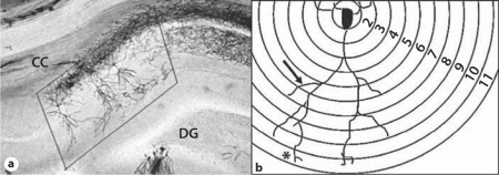
The hippocampus develops rapidly during the late fetal and early postnatal periods. Fetal/neonatal iron deficiency anemia (IDA) alters the genomic expression, neurometabolism and electrophysiology of the hippocampus during the period of IDA and, strikingly, in adulthood despite neonatal iron treatment. To determine how early IDA affects the structural development of the apical dendrite arbor in hippocampal area CA1 in the offspring, pregnant rat dams were given an iron-deficient (ID) diet between gestational day 2 and postnatal day (P) 7 followed by rescue with an iron-sufficient (IS) diet. Apical dendrite morphology in hippocampus area CA1 was assessed at P15, P30 and P70 by Scholl analysis of Golgi-Cox-stained neurons. Messenger RNA levels of nine cytoplasmic and transmembrane proteins that are critical for dendrite growth were analyzed at P7, P15, P30 and P65 by quantitative real-time polymerase chain reaction. The ID group had reduced transcript levels of proteins that modify actin and tubulin dynamics [e.g. cofilin-1 (Cfl-1), profilin-1 (Pfn-1), and profilin-2 (Pfn-2)] at P7, followed at P15 by a proximal shift in peak branching, thinner third-generation dendritic branches and smaller-diameter spine heads. At P30, iron treatment since P7 resulted in recovery of all transcripts and structural components except for a continued proximal shift in peak branching. Nevertheless, at P65-P70, the formerly ID group showed a 32% reduction in 9 mRNA transcripts, including Cfl-1 and Pfn-1 and Pfn-2, accompanied by 25% fewer branches, that were also proximally shifted. These alterations may be due to early-life programming of genes important for structural plasticity during adulthood and may contribute to the abnormal long-term electrophysiology and recognition memory behavior that follows early iron deficiency.
Copyright 2010 S. Karger AG, Basel.
Figures


References
-
- Bagot RC, van Hasselt FN, Champagne DL, Meaney MJ, Krugers HJ, Joels M. Maternal care determines rapid effects of stress mediators on synaptic plasticity in adult rat hippocampal dentate gyrus. Neurobiol Learn Mem. 2009;92:292–300. - PubMed
-
- Champagne DL, Bagot RC, van Hasselt F, Ramakers G, Meaney MJ, de Kloet ER, Joels M, Krugers H. Maternal care and hippocampal plasticity: evidence for experience-dependent structural plasticity, altered synaptic functioning, and differential responsiveness to glucocorticoids and stress. J Neurosci. 2008;28:6037–6045. - PMC - PubMed
-
- Williams CL. Food for thought: brain, genes, and nutrition. Brain Res. 2008;1237:1–4. - PubMed
-
- Siddappa AM, Georgieff MK, Wewerka S, Worwa C, Nelson CA, Deregnier RA. Iron deficiency alters auditory recognition memory in newborn infants of diabetic mothers. Pediatr Res. 2004;55:1034–1041. - PubMed
MeSH terms
Substances
Grants and funding
LinkOut - more resources
Full Text Sources
Other Literature Sources
Miscellaneous
