

Review
doi: 10.3858/emm.2010.42.9.064.
Molecular imaging of membrane proteins and microfilaments using atomic force microscopy
Affiliations
- PMID: 20689364
- PMCID: PMC2947017
- DOI: 10.3858/emm.2010.42.9.064
Item in Clipboard
Review
Molecular imaging of membrane proteins and microfilaments using atomic force microscopy
Exp Mol Med.
.
Abstract
Atomic force microscopy (AFM) is an emerging technique for a variety of uses involving the analysis of cells. AFM is widely applied to obtain information about both cellular structural and subcellular events. In particular, a variety of investigations into membrane proteins and microfilaments were performed with AFM. Here, we introduce applications of AFM to molecular imaging of membrane proteins, and various approaches for observation and identification of intracellular microfilaments at the molecular level. These approaches can contribute to many applications of AFM in cell imaging.
Figures
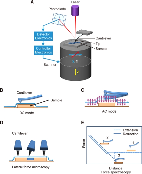
Principle of operation and operation modes in AFM. (A) Schematics of atomic force microscopy principle. (B-E) Four widely used atomic force microscopy operation modes: (B) DC mode, (C) AC mode, (D) lateral force microscopy, and (E) Force spectroscopy. A typical force-distance curve is shown (E). The cantilever is not deflected yet (1). As the tip approaches to the surface, the cantilever bends upward by repulsive forces (2). As the tip retracts from the surface, the cantilever bends downward by attractive forces between the tip and sample (3).

Identification of microfilaments by on-stage labeling/imaging. Samples mounted on the scanner tube are sequentially imaged in aqueous solution by AFM before (pre-imaging) and after (post-labeling) labeling with molecular probes including antibodies. Line profiles from pre-imaging (from a to b) and post-imaging (from a' to b') are compared.
Similar articles
-
High-speed atomic force microscopy: imaging and force spectroscopy.FEBS Lett. 2014 Oct 1;588(19):3631-8. doi: 10.1016/j.febslet.2014.06.028. Epub 2014 Jun 14. FEBS Lett. 2014. PMID: 24937145 Review.
-
Imaging fibroblast cells using atomic force microscopy (AFM).Cold Spring Harb Protoc. 2010 Oct 1;2010(10):pdb.prot5500. doi: 10.1101/pdb.prot5500. Cold Spring Harb Protoc. 2010. PMID: 20889697
-
Identification and ultrastructural imaging of photodynamic therapy-induced microfilaments by atomic force microscopy.Ultramicroscopy. 2009 Nov;109(12):1428-34. doi: 10.1016/j.ultramic.2009.07.009. Epub 2009 Jul 24. Ultramicroscopy. 2009. PMID: 19665305
-
Cytoskeleton induced the changes of microvilli and mechanical properties in living cells by atomic force microscopy.J Cell Physiol. 2021 May;236(5):3725-3733. doi: 10.1002/jcp.30110. Epub 2020 Nov 10. J Cell Physiol. 2021. PMID: 33169846
-
Nanoscale imaging and force probing of biomolecular systems using atomic force microscopy: from single molecules to living cells.Nanoscale. 2017 Nov 23;9(45):17643-17666. doi: 10.1039/c7nr07023c. Nanoscale. 2017. PMID: 29135007 Review.
Cited by
-
Investigating cell mechanics with atomic force microscopy.J R Soc Interface. 2015 Mar 6;12(104):20140970. doi: 10.1098/rsif.2014.0970. J R Soc Interface. 2015. PMID: 25589563 Free PMC article.
-
Dysferlin-peptides reallocate mutated dysferlin thereby restoring function.PLoS One. 2012;7(11):e49603. doi: 10.1371/journal.pone.0049603. Epub 2012 Nov 20. PLoS One. 2012. PMID: 23185377 Free PMC article.
-
Targeting cell-matrix interface mechanobiology by integrating AFM with fluorescence microscopy.Prog Biophys Mol Biol. 2022 Dec;176:67-81. doi: 10.1016/j.pbiomolbio.2022.08.005. Epub 2022 Aug 30. Prog Biophys Mol Biol. 2022. PMID: 36055517 Free PMC article. Review.
-
Measurement of cationic and intracellular modulation of integrin binding affinity by AFM-based nanorobot.Biophys J. 2013 Jul 2;105(1):40-7. doi: 10.1016/j.bpj.2013.05.052. Biophys J. 2013. PMID: 23823222 Free PMC article.
-
Understanding nanoparticle endocytosis to improve targeting strategies in nanomedicine.Chem Soc Rev. 2021 May 7;50(9):5397-5434. doi: 10.1039/d0cs01127d. Epub 2021 Mar 5. Chem Soc Rev. 2021. PMID: 33666625 Free PMC article. Review.
References
-
- Almeida RD, Manadas BJ, Carvalho AP, Duarte CB. Intracellular signaling mechanisms in photodynamic therapy. Biochim Biophys Acta. 2004;1704:59–86. - PubMed
-
- Ando T, Uchihashi T, Kodera N, Miyagi A, Nakakita R, Yamashita H, Sakashita M. High-speed atomic force microscopy for studying the dynamic behavior of protein molecules at work. Jpn J Appl Phys. 2008a;45:1897–1903.
-
- Ando T, Uchihashi T, Kodera N, Yamamoto D, Miyagi A, Taniguchi M, Yamashita H. High-speed AFM and nano-visualization of biomolecular processes. Pflugers Arch. 2008b;456:211–225. - PubMed
-
- Azoury J, Verlhac MH, Dumont J. Actin filaments: key players in the control of asymmetric divisions in mouse oocytes. Biol Cell. 2009;101:69–76. - PubMed
Publication types
MeSH terms
Substances
LinkOut - more resources
Full Text Sources
Miscellaneous