. 2011 Feb;18(2):282-92.
doi: 10.1038/cdd.2010.92. Epub 2010 Aug 6.

Bid-mediated mitochondrial damage is a key mechanism in glutamate-induced oxidative stress and AIF-dependent cell death in immortalized HT-22 hippocampal neurons

Affiliations

Bid-mediated mitochondrial damage is a key mechanism in glutamate-induced oxidative stress and AIF-dependent cell death in immortalized HT-22 hippocampal neurons

S Tobaben et al. Cell Death Differ. 2011 Feb.

Abstract

Glutamate toxicity involves increases in intracellular calcium levels and enhanced formation of reactive oxygen species (ROS) causing neuronal dysfunction and death in acute and chronic neurodegenerative disorders. The molecular mechanisms mediating glutamate-induced ROS formation are, however, still poorly defined. Using a model system that lacks glutamate-operated calcium channels, we demonstrate that glutamate-induced acceleration of ROS levels occurs in two steps and is initiated by lipoxygenases (LOXs) and then significantly accelerated through Bid-dependent mitochondrial damage. The Bid-mediated secondary boost of ROS formation downstream of LOX activity further involves mitochondrial fragmentation and release of mitochondrial apoptosis-inducing factor (AIF) to the nucleus. These data imply that the activation of Bid is an essential step in amplifying glutamate-induced formation of lipid peroxides to irreversible mitochondrial damage associated with further enhanced free radical formation and AIF-dependent execution of cell death.


Figures

Figure 1
Figure 1
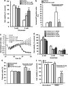
Glutamate leads to a time-dependent damage in HT-22 cells. (a) HT-22 cells were seeded in 96-well E-plates with a density of 4500 cells/well. Cells were observed for 48 h after seeding and treated with glutamate 5 mM after 24 h. Cell death became obvious 9–10 h after glutamate challenge. After onset of cell death HT-22 cells died within 3–5 h (n=8). (b) Cells were treated with glutamate at concentrations of 5 mM for 6, 12, 17 and 18 h. Cell death was detected by MTT assay (n=8). (c) Glutamate induces the production of lipid peroxides. Glutamate treatment was performed for 6–17 h. After addition of 2 μM BODIPY 581/591 C11 for 60 min, quantification was done by FACS analysis. Following glutamate exposure, a twofold increase in lipid peroxides after 6–8 h, and a secondary boost could be detected after 18 h (n=3). (d) Glutamate leads to formation of ROS and supports the production of lipid peroxides. Glutamate was added to the cells at a concentration of 3 mM. ROS levels were detected by dichlorodihydrofluorescein-diacetate (DCF) and quantified by FACS analysis (n=3). (ad) All experiments were repeated at least three times and results are reported as mean±S.D.
Figure 2
Figure 2
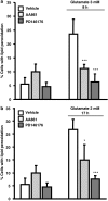
LOX inhibitors prevent peroxide formation in HT cells exposed to glutamate. Lipid peroxidation was detected 8 h (a) and up to 17 h (b) after onset of glutamate exposure by FACS analysis after staining of cells with BODIPY C11 (Ex=488 nm, Em=530 nm, 613 nm). The LOX inhibitor AA861 (0.1 μM) or PD146176 (0.5 μM) was added 1 h before the glutamate challenge (n=3). (a, b) All experiments were repeated three times and the results represent the mean ± S.D. *P<0.05, ***P<0.001 compared with glutamate-treated cells (ANOVA, Scheffë-test)
Figure 3
Figure 3
LOX inhibitors protect HT-22 cells against glutamate-induced cell death. (a) The LOX inhibitor PD146176 was applied 1 h before exposure to glutamate (5 mM) at concentrations of 0.1 and 0.5 μM. MTT (n=8) assay and FACS analysis were used to determine cell viability 18 h after onset of glutamate treatment. (b) LOX inhibitor PD146176 reduced the number of annexin-V/propidium iodide-positive cells compared with glutamate treated cells significantly. Cells were pretreated with the 12/15-LOX inhibitor PD146176 (0.5 μM) 1 h before glutamate challenge (5 mM). Cells were stained with annexin-V and propidium iodide and detected with FACS analysis (n=4). (c) HT-22 cells were seeded in 96-well E-plates with a density of 4500 cells/well. Cells were pretreated with PD146176 for 1 h and treated with glutamate 5 mM after 24 h (n=8). (d) The COX inhibitor indomethacin does not protect HT-22 cells against cell death induced by glutamate treatment. Indomethacin (5–100 μM) was added 1 h before exposure to glutamate at indicated concentrations. MTT assay was used to determine cell viability 18 h after glutamate treatment (n=8). (e) PD146176 protects primary cortical neurons against glutamate-induced cell death. Primary cortical neurons were pretreated with PD146176 0.5 μM 1 h before adding glutamate (20 μM). After 18 h cells were fixed with paraformaldehyde (4%) and stained with DAPI. The percentage of pyknotic nuclei was evaluated by counting 200 living and pyknotic cells per cell culture dish (n=5). (f) Primary cortical neurons were prepared from Alox-15 and wild-type mice at embryonic day E16. Cells were treated on day 6 in culture with glutamate (20 μM). Cell death was significantly reduced in Alox15 cultures compared with wild-type neurons. Cell death was evaluated by counting 200 DAPI-stained cells per dish (n=5). All data are provided as mean±S.D. **P<0.01, ***P<0.001 compared with glutamate-treated cells and ##P<0.01 compared with glutamate-treated wild-type neurons (ANOVA, Scheffé test)
Figure 4
Figure 4
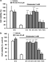
(a) LOX inhibitor PD146176 protects HT-22 cells when applied up to 8 h after glutamate treatment. The Lox inhibitor PD146176 was added at time points between 2 and 15 h after onset of glutamate treatment (5 mM). MTT assay was used to determine cell viability 18 h after onset of glutamate exposure (n=8). (b) Trolox protects HT-22 cells against glutamate toxicity when applied up to 8 h after glutamate challenge. Cells were treated with Trolox (50 μM) at time points between 2 and 15 h after glutamate (5 mM) challenge. Cell viability was detected by MTT assay (n=8). (a, b) All experiments were repeated three times and the results indicate the mean±S.D. ***P<0.001 compared with glutamate-treated cells (ANOVA, Scheffé-test)
Figure 5
Figure 5
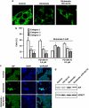
LOX inhibitor PD146176 0.5 μM prevents mitochondrial fragmentation and AIF-translocation to the nucleus. (a) HT-22 cells were transfected with mitoGFP using Lipofectamine 2000. After 24 h, the cells were damaged with glutamate 5 mM (n=4). (b) Mitochondrial morphology was analyzed by fluorescence microscopy and classified into three categories indicating the status of fission and fusion (category 1: tubulin-like, category 2: intermediate, category 3: fragmented) (n=4). (c) HT-22 cells were treated with glutamate 5 mM 24 h after seeding. PD146176 (0.5 μM) was applied 1 h before glutamate. Cells were fixed and immunostained 18 h after the treatment. Nuclei were stained with DAPI. Pictures were taken with a confocal microscope. The 12/15-LOX inhibitor prevented translocation of AIF to the nucleus. (d) HT-22 cells were damaged with glutamate and treated with PD146176 as indicated. After 14 h nuclear extracts were obtained for western blot analyses of AIF. HDAC1 served as a loading control (n=3). (ad) All experiments were repeated at least three times and all data are provided as mean ± S.D. *P<0.05, **P<0.01, ***P<0.001 compared with glutamate-treated cells (ANOVA, Scheffé test)
Figure 6
Figure 6
Bid inhibitor BI-6C9 protects cells against glutamate toxicity and prevents the secondary boost of ROS formation. Lipid peroxidation was detected 6–8 h (a) and up to 17 h (b) after onset of glutamate exposure by FACS analysis after staining cells with BODIPY C11 (Ex = 488 nm, Em = 530 and 613 nm). The Bid inhibitor BI-6C9 was present in the medium 1 h before and during the glutamate challenge (n=3). (c) HT-22 cells were damaged with glutamate (5 mM) and BI-6C9 (10 μM). Cell viability was detected by MTT assay 18 h after the onset of glutamate treatment (n=8). All experiments were repeated three times and the results indicate the mean ± S.D. ***P<0.001compared with glutamate-treated cells (ANOVA, Scheffé test). (d) Glutamate does not induce Bid cleavage in HT-22 cells. HT-22 cells were treated with glutamate for 2–18 h. To test whether the anti-Bid antibody could detect full-length Bid as well as tBid, control cell extracts were incubated with recombinant caspase-8 in vitro before performing the indicated immunoblot detection (upper panel). In contrast to caspase-8-incubated cell extracts, protein extracts from HT-22 cells exposed to glutamate did not reveal Bid cleavage at the indicated time points (lower panel)
Figure 7
Figure 7
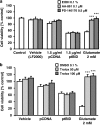
LOX inhibitors or Trolox does not attenuate the cytotoxic effect of tBid. (a) PD146176 (0.5 μM) and AA861 (0.1 μM) were added to HT-22 cells before transfection with tBid. Cell death was detected by using MTT assay 14 h after the transfection (n=3). (b) The vitamin E analog Trolox (50, 100 μM) was applied before transfection of HT-22 cells with tBid or exposure to glutamate. MTT assay was used to determine cell viability 24 h later (n=3). (a, b) All experiments were repeated three times and the results are reported as mean ± S.D. ***P<0.001 compared with glutamate-treated cells (ANOVA, Scheffé test)
Figure 8
Figure 8
Overview on the mechanisms of glutamate-induced cell death in HT-22 cells. Glutamate induces glutathione depletion by inhibition of xc-transporters and consequently leads to GpX4 depletion and increased 12/15-lipoxygenase activity followed by an increase in lipid peroxides. This leads to Bid activation and mitochondrial damage followed by a second boost in ROS production and release of mitochondrial pro-apoptotic proteins such as AIF

Similar articles

Cited by

References

    1. Culmsee C, Krieglstein J. Emerging pharmacotherapeutic strategies for the treatment of ischemic stroke. Drug Discov Today Ther Strateg. 2006;3:621–638.
    1. Lo EH, Dalkara T, Moskowitz MA. Mechanisms, challenges and opportunities in stroke. Nat Rev Neurosci. 2003;4:399–415. - PubMed
    1. Culmsee C, Junker V, Kremers W, Thal S, Plesnila N, Krieglstein J. Combination therapy in ischemic stroke: synergistic neuroprotective effects of memantine and clenbuterol. Stroke. 2004;35:1197–1202. - PubMed
    1. Fisher M, Schaebitz W. An overview of acute stroke therapy: past, present, and future. Arch Intern Med. 2000;160:3196–3206. - PubMed
    1. Matsuda S, Umeda M, Uchida H, Kato H, Araki T. Alterations of oxidative stress markers and apoptosis markers in the striatum after transient focal cerebral ischemia in rats. J Neural Transm. 2009;116:395–404. - PubMed

Publication types

MeSH terms