

The chemopreventive effects of Protandim: modulation of p53 mitochondrial translocation and apoptosis during skin carcinogenesis
- PMID: 20689586
- PMCID: PMC2912769
- DOI: 10.1371/journal.pone.0011902
The chemopreventive effects of Protandim: modulation of p53 mitochondrial translocation and apoptosis during skin carcinogenesis
Abstract
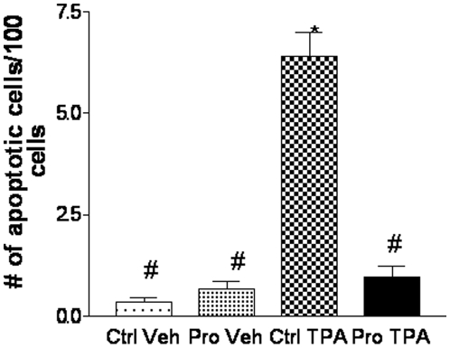
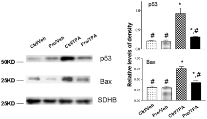
Protandim, a well defined dietary combination of 5 well-established medicinal plants, is known to induce endogenous antioxidant enzymes, such as manganese superoxide dismutase (MnSOD). Our previous studies have shown through the induction of various antioxidant enzymes, products of oxidative damage can be decreased. In addition, we have shown that tumor multiplicity and incidence can be decreased through the dietary administration of Protandim in the two-stage skin carcinogenesis mouse model. It has been demonstrated that cell proliferation is accommodated by cell death during DMBA/TPA treatment in the two-stage skin carcinogenesis model. Therefore, we investigated the effects of the Protandim diet on apoptosis; and proposed a novel mechanism of chemoprevention utilized by the Protandim dietary combination. Interestingly, Protandim suppressed DMBA/TPA induced cutaneous apoptosis. Recently, more attention has been focused on transcription-independent mechanisms of the tumor suppressor, p53, that mediate apoptosis. It is known that cytoplasmic p53 rapidly translocates to the mitochondria in response to pro-apoptotic stress. Our results showed that Protandim suppressed the mitochondrial translocation of p53 and mitochondrial outer membrane proteins such as Bax. We examined the levels of p53 and MnSOD expression/activity in murine skin JB6 promotion sensitive (P+) and promotion-resistant (P-) epidermal cells. Interestingly, p53 was induced only in P+ cells, not P- cells; whereas MnSOD is highly expressed in P- cells when compared to P+ cells. In addition, wild-type p53 was transfected into JB6 P- cells. We found that the introduction of wild-type p53 promoted transformation in JB6 P- cells. Our results suggest that suppression of p53 and induction of MnSOD may play an important role in the tumor suppressive activity of Protandim.
Conflict of interest statement
Figures


Similar articles
-
Manganese superoxide dismutase deficiency enhances cell turnover via tumor promoter-induced alterations in AP-1 and p53-mediated pathways in a skin cancer model.Oncogene. 2002 May 30;21(24):3836-46. doi: 10.1038/sj.onc.1205477. Oncogene. 2002. PMID: 12032821
-
Protandim, a fundamentally new antioxidant approach in chemoprevention using mouse two-stage skin carcinogenesis as a model.PLoS One. 2009;4(4):e5284. doi: 10.1371/journal.pone.0005284. Epub 2009 Apr 22. PLoS One. 2009. PMID: 19384424 Free PMC article.
-
p53 translocation to mitochondria precedes its nuclear translocation and targets mitochondrial oxidative defense protein-manganese superoxide dismutase.Cancer Res. 2005 May 1;65(9):3745-50. doi: 10.1158/0008-5472.CAN-04-3835. Cancer Res. 2005. PMID: 15867370
-
Manganese superoxide dismutase: beyond life and death.Amino Acids. 2012 Jan;42(1):139-58. doi: 10.1007/s00726-010-0600-9. Epub 2010 May 8. Amino Acids. 2012. PMID: 20454814 Free PMC article. Review.
-
Manganese superoxide dismutase vs. p53: regulation of mitochondrial ROS.Mitochondrion. 2010 Nov;10(6):649-61. doi: 10.1016/j.mito.2010.06.003. Epub 2010 Jul 1. Mitochondrion. 2010. PMID: 20601193 Review.
Cited by
-
Isocitrate dehydrogenase 1 is downregulated during early skin tumorigenesis which can be inhibited by overexpression of manganese superoxide dismutase.Cancer Sci. 2012 Aug;103(8):1429-33. doi: 10.1111/j.1349-7006.2012.02317.x. Epub 2012 Jun 7. Cancer Sci. 2012. PMID: 22533343 Free PMC article.
-
Antioxidant and Anti-Tumor Effects of Dietary Vitamins A, C, and E.Antioxidants (Basel). 2023 Mar 3;12(3):632. doi: 10.3390/antiox12030632. Antioxidants (Basel). 2023. PMID: 36978880 Free PMC article. Review.
-
The role of manganese superoxide dismutase in skin cancer.Enzyme Res. 2011;2011:409295. doi: 10.4061/2011/409295. Epub 2011 Mar 23. Enzyme Res. 2011. PMID: 21603266 Free PMC article.
-
Solanum lyratum Extracts Induce Extrinsic and Intrinsic Pathways of Apoptosis in WEHI-3 Murine Leukemia Cells and Inhibit Allograft Tumor.Evid Based Complement Alternat Med. 2012;2012:254960. doi: 10.1155/2012/254960. Epub 2012 May 7. Evid Based Complement Alternat Med. 2012. PMID: 22611426 Free PMC article.
-
Phloretin induces apoptosis of human esophageal cancer via a mitochondria-dependent pathway.Oncol Lett. 2017 Dec;14(6):6763-6768. doi: 10.3892/ol.2017.7037. Epub 2017 Sep 22. Oncol Lett. 2017. PMID: 29151915 Free PMC article.
References
-
- Zhao Y, Chaiswing L, Velez JM, Batinic-Haberle I, Colburn NH, et al. p53 translocation to mitochondria precedes its nuclear translocation and targets mitochondrial oxidative defense protein-manganese superoxide dismutase. Cancer Res. 2005;65:3745–3750. - PubMed
-
- Bowden GT, Finch J, Domann F, Krieg P. Molecular mechanisms involved in skin tumor initiation, promotion, and progression. 1995. pp. 99–111. CRC Press, Inc.
-
- Avila GE, Zheng X, Cui XX, Ryan AD, Hansson A, et al. Inhibitory effects of 12-O-tetradecanoylphorbol-13-acetate alone or in combination with all-trans retinoic acid on the growth of cultured human pancreas cancer cells and pancreas tumor xenographs in immunodeficient mice. J Pharmacol Exp Ther. 2005;15:170–187. - PubMed
-
- Zheng X, Chang RL, Cui XX, Avila GE, Hebbar V, et al. Effects of 12-O-tetradecanoylphorbol-13-acetate (TPA) in combination with paclitaxel (Taxol) on prostate cancer LnCap cells cultured in vitro or grown as xenongraft tumors in immunodeficient mice. Clin Cancer Res. 2006;1:3444–3451. - PubMed
-
- Zhang X, Li W, Olumi AF. Low dose 12-O-tetradecanoylphorbol-13-acetate enhances tumor necrosis factor related apoptosis-inducing ligand induced apoptosis in prostate cancer cells. Clin Cancer Res. 2007;13:7181–7190. - PubMed
Publication types
MeSH terms
Substances
LinkOut - more resources
Full Text Sources
Medical
Research Materials
Miscellaneous
