. 2010 Jul 30;5(7):e11908.
doi: 10.1371/journal.pone.0011908.

Epstein-Barr virus interferes with the amplification of IFNalpha secretion by activating suppressor of cytokine signaling 3 in primary human monocytes

Affiliations

Epstein-Barr virus interferes with the amplification of IFNalpha secretion by activating suppressor of cytokine signaling 3 in primary human monocytes

François Michaud et al. PLoS One. .

Abstract

Background: Epstein-Barr virus is recognized to cause lymphoproliferative disorders and is also associated with cancer. Evidence suggests that monocytes are likely to be involved in EBV pathogenesis, especially due to a number of cellular functions altered in EBV-infected monocytes, a process that may affect efficient host defense. Because type I interferons (IFNs) are crucial mediators of host defense against viruses, we investigated the effect of EBV infection on the IFNalpha pathway in primary human monocytes.

Methodology/principal findings: Infection of monocytes with EBV induced IFNalpha secretion but inhibited the positive feedback loop for the amplification of IFNalpha. We showed that EBV infection induced the expression of suppressor of cytokine signaling 3 (SOCS3) and, to a lesser extent, SOCS1, two proteins known to interfere with the amplification of IFNalpha secretion mediated by the JAK/STAT signal transduction pathway. EBV infection correlated with a blockage in the activation of JAK/STAT pathway members and affected the level of phosphorylated IFN regulatory factor 7 (IRF7). Depletion of SOCS3, but not SOCS1, by small interfering RNA (siRNA) abrogated the inhibitory effect of EBV on JAK/STAT pathway activation and significantly restored IFNalpha secretion. Finally, transfection of monocytes with the viral protein Zta caused the upregulation of SOCS3, an event that could not be recapitulated with mutated Zta.

Conclusions/significance: We propose that EBV protein Zta activates SOCS3 protein as an immune escape mechanism that both suppresses optimal IFNalpha secretion by human monocytes and favors a state of type I IFN irresponsiveness in these cells. This immunomodulatory effect is important to better understand the aspects of the immune response to EBV.


Conflict of interest statement

Competing Interests: The authors have declared that no competing interests exist.

Figures

Figure 1
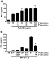
Figure 1. Effect of EBV infection on IFNα secretion by human monocytes.
Monocytes (2×10⁶ cells) were stimulated with (A) poly(I:C) or (B) EBV for 20 hours. Following stimulation, cell-free supernatants were harvested (1st stimulation). Cells were restimulated a second time with poly(I:C) or EBV for another 20 hours (2nd stimulation) and cell-free supernatants were harvested for IFNα determination by ELISA. Data are representative of three independent experiments. *p≤0.05 when compared to EBV 1st stimulation.
Figure 2
Figure 2. Expression of SOCS proteins following EBV infection of human monocytes.
Monocytes (5×10⁶) were stimulated or not with EBV for the indicated time. (A) The expression of SOCS1 and SOCS3 proteins was evaluated by RT-PCR using described primers (Table 1) or (B) by Western blot analysis using specific anti-SOCS1 antibodies, anti-SOCS3 antibodies and anti-Actin as loading control. Densitometry was performed and represents fold protein induction (relative to 0 min.) ± std. dev. of experiments performed in duplicate. Data are representative of three independent experiments.
Figure 3
Figure 3. Effect of EBV infection on the activation of the JAK/STAT pathway.
(A) Monocytes were incubated in the presence of IFNα (1000 U/ml) for 15 minutes and 20 hours. Following incubation (20 hours), cells were restimulated or not with IFNα for 15 minutes. The expression of phospho(p)STAT1, and phospho(p)STAT2 proteins was evaluated by Western blot analysis. Membranes were also probed with anti-Actin as a loading control. Densitometry was performed and represents fold protein induction (relative to non stimulated cells) ± std. dev. of experiments performed in duplicate. (B) Monocytes (5×10⁶) were treated with IFNα (1000 U/ml) for 15 minutes, with EBV for 20 hours or were pre-incubated for 20 hours in the presence of EBV followed by a stimulation with IFNα for 15 minutes. The expression of phospho(p)Tyk2, phospho(p)JAK1, phospho(p)STAT1, and phospho(p)STAT2 proteins was evaluated by Western blot analysis. Membranes were also probed with anti-Tyk2, JAK1, STAT1 and STAT2 as a loading control. (C) Monocytes (2×10⁶ cells) were transfected with 165 nM siRNA targeting SOCS1 or SOCS3 prior to EBV stimulation for 1 hour. Scramble siRNA was used as control. The expression of SOCS1 and SOCS3 was evaluated by Western blot analysis. Densitometry was performed and represents fold protein induction (relative to non-transfected cells) ± std. dev. of experiments performed in duplicate. (D) Monocytes (2×10⁶ cells) were either left untransfected or were transfected with siRNA targeting SOCS1 or SOCS3 and stimulated as in (B). The expression of phospho(p)STAT1 and phospho(p)STAT2 proteins was evaluated by Western blot analysis. Membranes were also probed with anti-STAT1 and STAT2 as a loading control. Data are representative of three independent experiments. NS: non-stimulated; NT: non-transfected; SCR: scrambled siRNA.
Figure 4
Figure 4. Effect of EBV infection on IRF3 and IRF7 activation.
1st EBV stimulation: Monocytes (5×10⁶ cells) were stimulated or not with EBV for the indicated times and expression of phosphorylated forms of IRF3 and IRF7 proteins was evaluated by Western blot analysis. 2nd EBV stimulation: Monocytes were first stimulated with EBV for 20 hours, washed and then restimulated a second time with EBV for the indicated times. Expression of phosphorylated forms of IRF3 and IRF7 proteins was evaluated by Western blot analysis. Membranes were also probed with anti-Actin as a loading control. Densitometry was performed and represents fold protein induction (relative to 0 h) ± std. dev. of experiments performed in duplicate. Data are representative of three independent experiments.
Figure 5
Figure 5. siRNA against SOCS3 restores IFNα secretion following EBV infection.
Monocytes (2×10⁶ cells) were transfected with 165 nM siRNA targeting SOCS1 or SOCS3. Scramble siRNA was used as control. Twenty-four hours after transfection, cells were stimulated with EBV for 20 hours (1st EBV stimulation). Following this first EBV stimulation, medium was replaced and cells were stimulated a second time with EBV (2nd EBV stimulation) for an additional 20 hours. Cell-free supernatants were then harvested for IFNα determination. Data are representative of three independent experiments. Values in percentage represent the inhibition in IFNα secretion relative to the first respective EBV stimulation. *p≤0.05 compared to cells transfected with scramble siRNA and stimulated a second time with EBV. SCR: scrambled siRNA.
Figure 6
Figure 6. Activation of SOCS3 by the viral protein Zta.
(A) Monocytes (5×10⁶) were mock- or EBV-stimulated for 20 hours and expression of the EBV protein Zta was evaluated by Western blot analysis. (B) HEK293 cells were transiently co-transfected with a vector encoding WT Zta or mutated Zta (ΔZta) at indicated concentrations along with a SOCS3 promoter-driven luciferase reporter vector. Luciferase assay was performed 48 hours post-transfection. (C) Monocytes (5×10⁶) were transfected with 300 ng of either Zta vector or ΔZta vector or mock vector control (CTRL). Forty-eight hours following transfection, monocytes were stimulated with IFNα (1000 U/ml) for 15 minutes. The expression of SOCS3 and phospho(p)STAT2 proteins was evaluated by Western blot analysis. Membranes were also probed with anti-Actin as a loading control. Densitometry was performed and represents fold protein induction (relative to mock-transfected cells) ± std. dev. of experiments performed in duplicate. Data are representative of two independent experiments. *p≤0.05 compared to non-transfected control cells. NS: non-stimulated; NT: non-transfected.
Figure 7
Figure 7. Proposed mechanism for EBV-mediated interference with type I IFN secretion by human monocytes.
Following entry into the cell, EBV induces IRF3/7 activation leading to the initial production of IFNα/β. EBV infection also induces the synthesis of SOCS3 protein which results in inhibition of IFN receptor (IFNR) signaling and also in inhibition of the amplification of IFNα/β production. ISGs: interferon-stimulated genes. Black lines indicate “activation” and red lines indicate “suppression”. Red dotted lines represent suppressive effects of SOCS protein activation on JAK/STAT signaling events.

Similar articles

Cited by

References

    1. Straus SE, Cohen JI, Tosato G, Meier J. NIH conference. Epstein-Barr virus infections: biology, pathogenesis, and management. Ann Intern Med. 1993;118:45–58. - PubMed
    1. Kasahara Y, Yachie A. Cell type specific infection of Epstein-Barr virus (EBV) in EBV-associated hemophagocytic lymphohistiocytosis and chronic active EBV infection. Crit Rev Oncol Hematol. 2002;44:283–294. - PubMed
    1. Savard M, Gosselin J. Epstein-Barr virus immunossuppression of innate immunity mediated by phagocytes. Virus Res. 2006;119:134–145. - PubMed
    1. Gosselin J, Menezes J, D'Addario M, Hiscott J, Flamand L, et al. Inhibition of tumor necrosis factor-alpha transcription by Epstein-Barr virus. Eur J Immunol. 1991;21:203–208. - PubMed
    1. Jabs WJ, Wagner HJ, Maurmann S, Hennig H, Kreft B. Inhibition of macrophage inflammatory protein-1 alpha production by Epstein-Barr virus. Blood. 2002;99:1512–1516. - PubMed

Publication types

MeSH terms

Substances