. 2010 Jul 30;5(7):e11921.
doi: 10.1371/journal.pone.0011921.

Burn injury reduces neutrophil directional migration speed in microfluidic devices

Affiliations

Burn injury reduces neutrophil directional migration speed in microfluidic devices

Kathryn L Butler et al. PLoS One. .

Abstract

Thermal injury triggers a fulminant inflammatory cascade that heralds shock, end-organ failure, and ultimately sepsis and death. Emerging evidence points to a critical role for the innate immune system, and several studies had documented concurrent impairment in neutrophil chemotaxis with these post-burn inflammatory changes. While a few studies suggest that a link between neutrophil motility and patient mortality might exist, so far, cumbersome assays have prohibited exploration of the prognostic and diagnostic significance of chemotaxis after burn injury. To address this need, we developed a microfluidic device that is simple to operate and allows for precise and robust measurements of chemotaxis speed and persistence characteristics at single-cell resolution. Using this assay, we established a reference set of migration speed values for neutrophils from healthy subjects. Comparisons with samples from burn patients revealed impaired directional migration speed starting as early as 24 hours after burn injury, reaching a minimum at 72-120 hours, correlated to the size of the burn injury and potentially serving as an early indicator for concurrent infections. Further characterization of neutrophil chemotaxis using this new assay may have important diagnostic implications not only for burn patients but also for patients afflicted by other diseases that compromise neutrophil functions.


Conflict of interest statement

Competing Interests: The authors have declared that no competing interests exist.

Figures

Figure 1
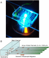
Figure 1. Overview of the neutrophil chemotaxis device.
a. Microfluidic devices mounted on glass slides are observed on a microscope stage. In this picture, six chemotaxis devices are aligned side by side on the same glass slide. The left most device has inlet and outlet tubing attached and is filled with green fluorescent dye. b. Schematic of the microfluidic device. The device consists of a larger main channel and an orthogonal array of smaller channels. In the first step, the whole device, including the side channels, is primed with the chemoattractant solution. In the second step, neutrophils suspended in buffer are introduced in the main channel, washing out the chemoattractant from the main channel. The chemoattractant gradient is established by diffusion, in the longitudinal direction of the side channels, between the end of the side channels filled with chemoattractant and the main channel filled with buffer. Neutrophils inside the main channel follow a chemoattractant gradient and enter an array of side channels, where their chemotaxis migration speed is measured.
Figure 2
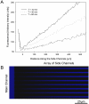
Figure 2. Visualization of the chemical gradients along the side channels.
a. Linear gradients, formed along the side channels, were imaged using fluorescent dyes of molecular weight comparable with that of the chemoattractant. Starting immediately after replacing the solution in the main channel with buffer, quantitative fluorescence was measured along the array of side channels at 10, 30 and 60 minutes. These measurements demonstrate the relative stability of the linear chemoattractant gradient over time. b. At 30 minutes after introducing the buffer, a linear gradient of fluorescent dye can be visualized along the side channels. The height of the main channel (to the left) is larger that that of the side channels and accounts for the apparent higher fluorescence of the main channel compared to the side channels.
Figure 3
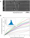
Figure 3. Neutrophil migration inside the array of channels.
a. Neutrophils enter the array of channels and move along the channels towards higher concentrations of chemoattractant – see also Movie S1. b. Displacement of neutrophils vs. time inside the array of channels. For the first 10 minutes after entering the side channels neutrophil display remarkably uniform migration speed. Insert shows the distribution of average speed of migration for 800 neutrophils from one healthy donor.
Figure 4
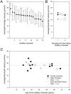
Figure 4. Neutrophil motility in healthy donors.
a. Average neutrophil motility in 18 healthy donors. Samples were rearranged in decreasing order of the average motilities. Bars represent standard error of the mean. The motility of at least 50 neutrophils was calculated for each sample. b. Validation of the repeatability of neutrophil motility. Two samples from the same healthy donors were collected at two weeks time interval and neutrophil migration speed measured in the microfluidic devices. c. Distribution of average values for neutrophil motility with the age of healthy donors. No significant changes in neutrophil motility were observed with increasing age of the healthy volunteers. No significant differences were observed between female and male donors (filled and empty dots, respectively).
Figure 5
Figure 5. Correlations between neutrophil motility and clinical parameters in burn patients.
a. We observed significant correlations between neutrophil motility at 72 hours after burn injury and total burn surface area (R2 = 0.6). b. No significant correlation was found between neutrophil motility and the temperature in burn patients (R2 = 0.2). Samples from the same patient are coded using the same symbol.
Figure 6
Figure 6. Correlations between neutrophil motility and neutrophil counts in burn patients.
a. No significant correlation was found between neutrophil motility and the absolute neutrophil count (R2 = 0.2). b. Also, there was no significant correlation between average neutrophil motility and percentage of band cells in the neutrophil population (R2 = 0.01).
Figure 7
Figure 7. Changes in neutrophil motility in burn patients.
Average neutrophil motility in individual patients was measured at 48 hours intervals after admission to the hospital. Lower values for neutrophil migration speed compared to healthy volunteers (dashed gray line) were recorded as early as 24 hours after burn injury for the majority of the patients. Insert shows the changes in average neutrophil migration speed for all patients, with respect to the time of injury.

Similar articles

Cited by

References

    1. White CE, Renz EM. Advances in surgical care: management of severe burn injury. Crit Care Med. 2008;36(7) Suppl:S318–324. - PubMed
    1. Salinas J, Drew G, Gallagher J, Cancio LC, Wolf SE, et al. Closed-loop and decision-assist resuscitation of burn patients. J Trauma. 2008;64(4) Suppl:S321–332. - PubMed
    1. Saffle JI. The phenomenon of "fluid creep" in acute burn resuscitation. J Burn Care Res. 2007;28(3):382–395. - PubMed
    1. Ipaktchi K, Arbabi S. Advances in burn critical care. Crit Care Med. 2006;34(9) Suppl:S239–244. - PubMed
    1. Ong YS, Samuel M, Song C. Meta-analysis of early excision of burns. Burns. 2006;32(2):145–150. - PubMed

Publication types