. 2010:2010:617052.
doi: 10.1155/2010/617052. Epub 2010 Jun 20.

Glucose regulation of thrombospondin and its role in the modulation of smooth muscle cell proliferation

Affiliations

Glucose regulation of thrombospondin and its role in the modulation of smooth muscle cell proliferation

Laura A Maile et al. Exp Diabetes Res. 2010.

Abstract

Smooth muscle cells (SMC) maintained in high glucose are more responsive to IGF-I than those in normal glucose. There is significantly more thrombospondin-1 (TSP-1) in extracellular matrix surrounding SMC grown in 25 mM glucose. In this study we investigated 1) the mechanism by which glucose regulates TSP-1 levels and 2) the mechanism by which TS-1 enhances IGF-I signaling. The addition of TSP-1 to primary SMC was sufficient to enhance IGF-I responsiveness in normal glucose. Reducing TSP-1 protein levels inhibited IGF-I signaling in SMC maintained in high glucose. We determined that TSP-1 protected IAP/CD47 from cleavage and thereby facilitated its association with SHP substrate-1 (SHPS-1). We have shown previously that the hyperglycemia induced protection of IAP from cleavage is an important component of the ability of hyperglycemia to enhance IGF-I signaling. Furthermore we determined that TSP-1 also enhanced phosphorylation of the beta3 subunit of the alphaVbeta3 integrin, another molecular event that we have shown are critical for SMC response to IGF-I in high glucose. Our studies also revealed that the difference in the amount of TSP-1 in the two different glucose conditions was due, at least in part, to a difference in the cellular uptake and degradation of TSP-1.


Figures

Figure 1
Figure 1
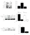
TSP-1 levels correlate with the level of intact IAP in SMC. (a) SMC were grown to confluency in DMEM containing 5 or 25 mM glucose and then incubated overnight in SFM with the appropriate level of glucose. Extracellular matrix, membrane extract or conditioned medium was harvested as described in the methods. Equal amounts of protein were immunoblotted (IB) with either an anti-TSP-1 or β3 antibody. (b) SMC were grown and incubated overnight in SFM as described for (a). The following day the incubation was continued for a further 6 hours after the addition (+) of TSP-1, (1 μg/mL). The graphs show arbitrary scanning units derived from the immunoblots with the anti-IAP antibody from three independent experiments. ***P < .005 when the level of IAP in the presence of TSP-1 is compared to SFM alone. (c) Equal amount of protein from the homogenates of aorta from normal (Con) and hyperglycemic (STZ) pigs were separated by SDS PAGE and the level of TSP-1, IAP and the IGF-IR determined by western immunoblotting (IB). The graphs shows show arbitrary scanning units derived from the immunoblots with the anti-TSP-1 or IAP antibody, as labeled, from three independent experiments. **P < .01 when the level of TSP-1/IAP from the STZ animals is compared with control. (d) SMC were grown and incubated overnight in SFM as described for (a). The following day the incubation was continued for a further 6 hours after the addition (+) of 4N1K or 4N1G, (1 μg/mL). The graph shows the fold difference in IAP levels compared with the level derived from SMC incubated in 25 mM glucose using arbitrary scanning units from the immunoblots with the anti-IAP antibody from three independent experiments.
Figure 2
Figure 2
The IAP binding domain of TSP-1 increases IGF-I signaling in normal glucose. (a) SMCs were grown to confluency in DMEM containing 5 mM glucose and then incubated overnight in SFM with 5 mM glucose. The following day the incubation was continued for 6 hours with (+) 4N1K or 4NG1G (1 μg/mL). Equal amounts of lysate were then subjected to immunoprecipitation (IP) with and anti-SHPS-1 antibody prior to immunoblotting (IB) with an anti-IAP antibody or immunoblotted directly with an anti-SHPS-1 antibody. The graph shows the results from three similar experiments expressed as arbitrary scanning units (***P < .005 when the amount of IAP associated with SHPS-1 is compared with the value for SMC incubated in 5 mM glucose containing medium). (b) SMCs were grown to confluency in DMEM containing 5 mM glucose and then incubated overnight in SFM with 5 mM glucose. The following day the incubation was continued for 6 hours with (+) 4N1K or 4NG1G (1 μg/mL) followed by IGF-I (50 ng/mL) for 5 minutes. Equal amounts of lysate were then subjected to immunoprecipitation (IP) with and anti-SHPS-1 antibody prior to immunoblotting (IB) with either an antiphosphotyrosine (p-Tyr) or Shc antibody or immunoblotted directly with an anti-SHPS-1 antibody. The graph shows the results from three similar experiments expressed as fold increase in response to IGF-I compared with no addition of peptide (***P < .005). (c) SMCs were grown to confluency in DMEM containing 5 mM glucose and then incubated overnight in SFM with 5 mM glucose. The following day the incubation was continued for 6 hours with (+) 4N1K or 4NG1G (1 μg/mL) followed by IGF-I (50 ng/mL) for 5 minutes. Equal amounts of lysate were then subjected to immunoblotting (IB) with either an anti-phosphoERK or total-ERK antibody. The graph shows the results from three similar experiments expressed as fold increase in response to IGF-I compared with no addition of peptide (***P < .005).
Figure 3
Figure 3
TSP-1 protein levels regulate the response of SMC to hyperglycemia. (a) SMCs expressing either the TSP-1 or a scrambled (Scr) RNAi construct were grown to confluency in DMEM containing 25 mM glucose and then incubated overnight in SFM with 25 mM. Equal amounts of lysates were either subject to immunoprecipitation (IP) followed by immunoblotting or immunoblotted (IB) directly with the antibody indicated. The graph shows the mean fold decrease of each protein in the RNAi cultures compared with the Scr control of three independent experiments, (***P < .005). (b) SMC expressing either the TSP-1 or a scrambled (Scr) RNAi construct were grown to confluency in DMEM containing 25 mM glucose and then incubated overnight in SFM with 25 mM prior to exposure to IGF-I (50 ng/mL) for 5 minutes (+). Equal amounts of lysates were then subject to immunoprecipitation (IP) followed by immunoblotting (IB) with the antibody indicated. The graph shows the mean fold increase in phosphorylation of each protein in response to IGF-I (***P < .005 when the extent of phosphorylation in the absence of IGF-I is compared to the addition of IGF-I). (c) SMC expressing either the TSP-1 or a scrambled (Scr) RNAi construct were grown to confluency in DMEM containing 25 mM glucose and then incubated overnight in SFM with 25 mM prior to exposure to IGF-I (50 ng/mL) for 5 minutes (+). Equal amounts of lysates were then subject to immunoblotting (IB) with either an antiphospho or total ERK antibody. The graph shows the mean fold increase in phosphorylation in response to IGF-I (***P < .005 when the extent of phosphorylation in the absence of IGF-I is compared to the addition of IGF-I).
Figure 4
Figure 4
Restoration of the IGF-I response by addition of the CD47/IAP binding domain of TSP-1. (a) SMCs expressing either the TSP-1 or a scrambled (Scr) RNAi construct were grown to confluency in DMEM containing 25 mM glucose and then incubated overnight in SFM with 25 mM glucose. After overnight incubation in SFM cells were incubated for 6 hours with 4N1K (1 μg/mL) prior to lysis. The amount of intact IAP in each lysate was determined by immunoblotting with the anti-IAP antibody specific for intact IAP and the association between IAP and SHPS-1 was determined by coimmunoprecipitation. The graphs shows the results derived from western immunoblots from three similar experiments expressed as arbitrary scanning units increase (***P < .005 when the amount in the TSP-1 lysate is compared with the Scr cell lysate). (b) SMCs expressing the TSP-1 RNAi construct were grown to confluency in DMEM containing 25 mM glucose and then incubated overnight in SFM with 25 mM glucose. After overnight incubation in SFM cells were incubated for 6 hours with 4N1K (1 μg/mL) then treated with IGF-I (+) where indicted for 5 minutes prior to lysis. SHPS-1 and Shc phosphorylation in equal amounts of cell lysate was determined by immunoprecipitation and immunoblotting with an antiphosphotyrosine antibody (PY99). The graphs shows the results derived from western immunoblots from three similar experiments expressed as fold increase in phosphorylation after treatment with IGF-I (***P < .005 when the phosphorylation in the presence of 4N1K is compared with the absence). (c) 2 × 10⁴ cells grown 25 mM were plated in each well of a 24 well plate prior to exposure to 4N1K (1 μg/mL) and IGF-I (100 ng/mL) in DMEM + 0.2% platelet poor plasma. 48 hours after the addition of IGF-I cell number was determined by trypan blue staining and counting. ***P < .005 when cell number in response to IGF-I is compared with control.
Figure 5
Figure 5
TSP-1 protein levels regulate β3 phosphorylation. SMCs expressing the TSP-1 or a scrambled (Scr) RNAi construct were grown to confluency in DMEM containing 25 mM glucose and then incubated overnight in SFM with 25 mM glucose prior to treatment with 4N1K or 4NGG (1 μg/mL) or TSP-1 (0.5 and 1 μg/mL) for 6 hours prior to lysis. The graph shows the mean fold decrease, of three independent experiments. ***P < .005 when the level in TSP-1 RNAi cultures is compared with controls.
Figure 6
Figure 6
Glucose regulation of TSP-1 mRNA and protein. (a) RNA prepared from whole cell lysates. One microgram of total RNA was reverse transcribed. Two μL of the cDNA was use in a RT-PCR reaction. A standard curve was generated using the 24 hour normal glucose sample (10-fold dilutions from 1 to 10,000). The amount of RNA is expressed relative to the level in the 24 hour normal glucose sample. (b) An equal number of SMCs were plated in growth medium containing 5 or 25 mM glucose. Extracellular matrix and cell lysates were prepared from parallel cultures after 24, 48, 72, 96, and 120 hours and equal amounts of protein were separated by SDS-PAGE and immunoblotted with the appropriate antibody. The graph show the results expressed as arbitrary scanning units, of three independent experiments. ***P < .005 when the level of TSP-1 protein at each time point is compared with the level at 24 hours.
Figure 7
Figure 7
TSP-1 degradation. (a)–(e) SMCs grown to confluency in medium containing 25 or 5 mM glucose were incubated overnight in SFM and then treated with (+) a cathepsin inhibitor (100 nM), (+) bafilomycin (100 nM) or vehicle (−) for 6 hours prior to extracellular matrix preparation and immunoblotting with the anti-TSP-1 antibody or cell lysis/immunoprecipitation with the antibodies indicated. The graphs show the results of arbitrary scanning units derived from the western immunoblots of three independent experiments. Vertical line indicates where discontinuous bands from the same gel were used.

Similar articles

Cited by

References

    1. Jones JI, Prevette T, Gockerman A, Clemmons DR. Ligand occupancy of the αVβ3 integrin is necessary for smooth muscle cells to migrate in response to insulin-like growth factor. Proceedings of the National Academy of Sciences of the United States of America. 1996;93(6):2482–2487. - PMC - PubMed
    1. Cercek B, Fishbein MC, Forrester JS, Helfant RH, Fagin JA. Induction of insulin-like growth factor I messenger RNA in rat aorta after balloon denudation. Circulation Research. 1990;66(6):1755–1760. - PubMed
    1. Hayry P, Myllarniemi M, Aavik E, et al. Stabile D-peptide analog of insulin-like growth factor-1 inhibits smooth muscle cell proliferation after carotid ballooning injury in the rat. FASEB Journal. 1995;9(13):1336–1344. - PubMed
    1. Maile LA, Capps BE, Ling Y, Xi G, Clemmons DR. Hyperglycemia alters the responsiveness of smooth muscle cells to insulin-like growth factor-I. Endocrinology. 2007;148(5):2435–2443. - PubMed
    1. Maile LA, Badley-Clarke J, Clemmons DR. The association between integrin-associated protein and SHPS-1 regulates insulin-like growth factor-I receptor signaling in vascular smooth muscle cells. Molecular Biology of the Cell. 2003;14(9):3519–3528. - PMC - PubMed

Publication types