

Prefrontal Cortex Modulation during Anticipation of Working Memory Demands as Revealed by Magnetoencephalography
- PMID: 20689717
- PMCID: PMC2906181
- DOI: 10.1155/2010/840416
Prefrontal Cortex Modulation during Anticipation of Working Memory Demands as Revealed by Magnetoencephalography
Abstract
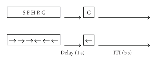
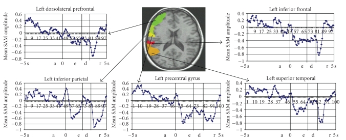
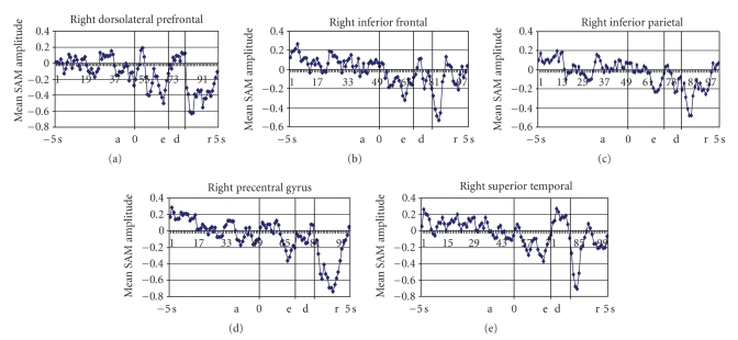
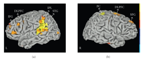
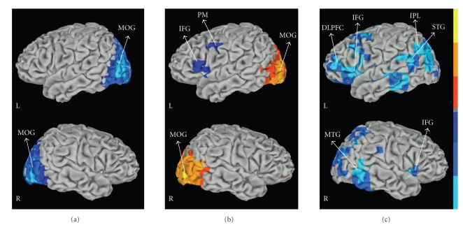
During the anticipation of task demands frontal control is involved in the assembly of stimulus-response mappings based on current goals. It is not clear whether prefrontal modulations occur in higher-order cortical regions, likely reflecting cognitive anticipation processes. The goal of this paper was to investigate prefrontal modulation during anticipation of upcoming working memory demands as revealed by magnetoencephalography (MEG). Twenty healthy volunteers underwent MEG while they performed a variation of the Sternberg Working Memory (WM) task. Beta band (14-30 Hz) SAM (Synthetic Aperture Magnetometry) analysis was performed. During the preparatory periods there was an increase in beta power (event-related synchronization) in dorsolateral prefrontal cortex (DLPFC) bilaterally, left inferior prefrontal gyrus, left parietal, and temporal areas. Our results provide support for the hypothesis that, during preparatory states, the prefrontal cortex is important for biasing higher order brain regions that are going to be engaged in the upcoming task.
Figures
Similar articles
-
Phase-Amplitude Coupling and Long-Range Phase Synchronization Reveal Frontotemporal Interactions during Visual Working Memory.J Neurosci. 2017 Jan 11;37(2):313-322. doi: 10.1523/JNEUROSCI.2130-16.2016. J Neurosci. 2017. PMID: 28077711 Free PMC article.
-
Oscillatory activity in bilateral prefrontal cortices is altered by distractor strength during working memory processing.Neuroimage. 2024 Nov 1;301:120878. doi: 10.1016/j.neuroimage.2024.120878. Epub 2024 Sep 30. Neuroimage. 2024. PMID: 39357689 Free PMC article.
-
Anodal Transcranial Direct Current Stimulation Induces High Gamma-Band Activity in the Left Dorsolateral Prefrontal Cortex During a Working Memory Task: A Double-Blind, Randomized, Crossover Study.Front Hum Neurosci. 2019 Apr 24;13:136. doi: 10.3389/fnhum.2019.00136. eCollection 2019. Front Hum Neurosci. 2019. PMID: 31105540 Free PMC article.
-
Cortical oscillatory power changes during auditory oddball task revealed by spatially filtered magnetoencephalography.Clin Neurophysiol. 2009 Mar;120(3):497-504. doi: 10.1016/j.clinph.2008.11.023. Epub 2009 Jan 12. Clin Neurophysiol. 2009. PMID: 19138878
-
Differential Effects of Transcranial Static Magnetic Stimulation Over Left and Right Dorsolateral Prefrontal Cortex on Brain Oscillatory Responses During a Working Memory Task.Neuroscience. 2023 May 1;517:50-60. doi: 10.1016/j.neuroscience.2023.03.006. Epub 2023 Mar 11. Neuroscience. 2023. PMID: 36907432
Cited by
-
Electrophysiological measures reveal the role of anterior cingulate cortex in learning from unreliable feedback.Cogn Affect Behav Neurosci. 2018 Oct;18(5):949-963. doi: 10.3758/s13415-018-0615-3. Cogn Affect Behav Neurosci. 2018. PMID: 29992483
-
The maintenance of complex visual scenes in working memory may require activation of working memory manipulation circuits in the dlPFC: a preliminary report.Mental Health Sci. 2024 Sep;2(3):e61. doi: 10.1002/mhs2.61. Epub 2024 May 10. Mental Health Sci. 2024. PMID: 39310119 Free PMC article.
-
Differential beta-band event-related desynchronization during categorical action sequence planning.PLoS One. 2013;8(3):e59544. doi: 10.1371/journal.pone.0059544. Epub 2013 Mar 18. PLoS One. 2013. PMID: 23527215 Free PMC article.
-
The impact of Val108/158Met polymorphism of catechol-O-methyltransferase on brain oscillations during working memory.Neurosci Lett. 2016 Jan 1;610:86-91. doi: 10.1016/j.neulet.2015.10.018. Epub 2015 Nov 1. Neurosci Lett. 2016. PMID: 26536074 Free PMC article.
-
Research on the MEG of Depression Patients Based on Multivariate Transfer Entropy.Comput Intell Neurosci. 2022 Jul 20;2022:7516627. doi: 10.1155/2022/7516627. eCollection 2022. Comput Intell Neurosci. 2022. PMID: 35909866 Free PMC article.
References
-
- Fuster JM. Prefrontal neurons in networks of executive memory. Brain Research Bulletin. 2000;52(5):331–336. - PubMed
-
- Sakai K, Passingham RE. Prefrontal interactions reflect future task operations. Nature Neuroscience. 2003;6(1):75–81. - PubMed
-
- De Pisapia N, Braver TS. Preparation for integration: the role of anterior prefrontal cortex in working memory. NeuroReport. 2008;19(1):15–19. - PubMed
-
- Lavric A, Mizon GA, Monsell S. Neurophysiological signature of effective anticipatory task-set control: a task-switching investigation. European Journal of Neuroscience. 2008;28(5):1016–1029. - PubMed
LinkOut - more resources
Full Text Sources
