

Choroidal pigmented lesions imaged by ultra-wide-field scanning laser ophthalmoscopy with two laser wavelengths (Optomap)
- PMID: 20689737
- PMCID: PMC2915871
- DOI: 10.2147/opth.s11864
Choroidal pigmented lesions imaged by ultra-wide-field scanning laser ophthalmoscopy with two laser wavelengths (Optomap)
Abstract
Purpose: Clinical differentiation of choroidal pigmented lesions is sometimes difficult. Choroidal melanoma is the most prevalent primary neoplasia among malignant ocular tumors, and metastasis often occurs before the primary tumor is diagnosed. Therefore, early detection is essential. We investigated the imaging properties of clinically diagnosed melanocytic choroidal tumors using a nonmydriatic ultra-wide-field scanning laser ophthalmoscope (SLO) with two laser wavelengths to distinguish benign from malignant lesions. Repeated standardized ultrasound (US) evaluation provided reference standard.
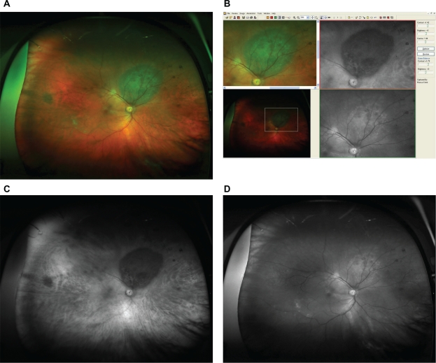
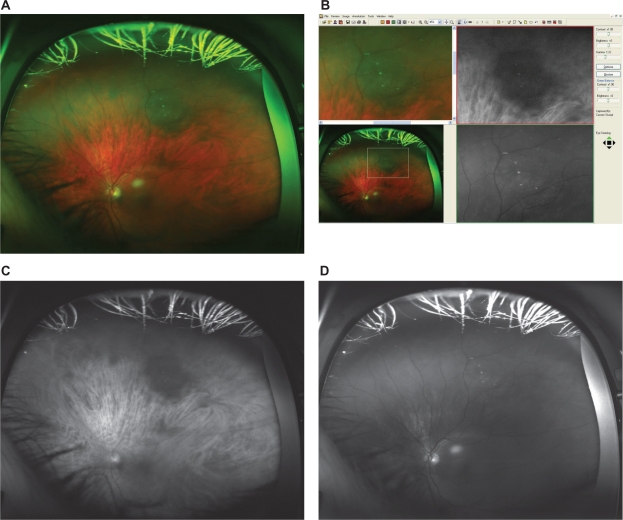
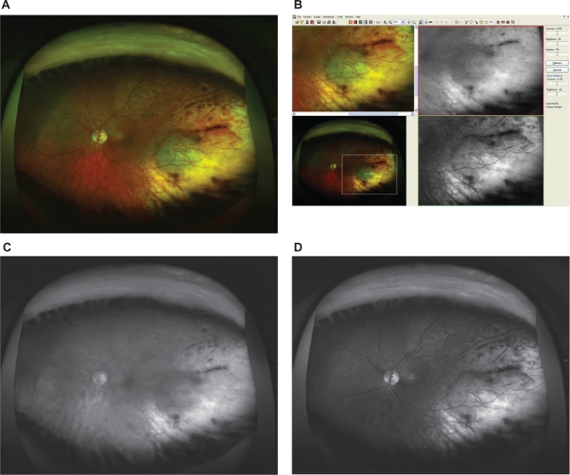
Methods: In a consecutive series of 49 patients with clinically diagnosed melanocytic choroidal tumors in one eye, 29 had established melanoma (defined by proven growth on repeated US follow-up) and 20 had nevi (defined by no malignancy according to clinical, US, and growth characteristics for at least 2 years). All patients underwent clinical examination, undilated Optomap((R)) (Optos PLC, Dunfermline, Fife, Scotland, UK) imaging, standardized US examination, and standard retinal photography. Measurements of the tumor base using the Optomap software were compared with US B-scan measurements. Imaging characteristics from the SLO images were correlated with the structural findings in the two patient groups.
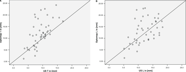
Results: Measurements of tumor base correlated well between SLO and US with r = 0.61 (T-direction) and r = 0.51 (L-direction). On SLO imaging, typical malignant lesions appeared dark on the red laser channel and bright on the green laser channel. Based on those simple binary characteristics, a sensitivity of 76% at a specificity of 70% was obtained for a correct classification of lesions. When analogous to clinical examination lesion size, margin touching the optic disc, and existence of subretinal fluid were additionally considered, 90% sensitivity at 82% specificity was obtained.
Conclusions: In this first, limited series, nonmydriatic SLO imaging with two laser wavelengths permitted to differentiate malignant ocular tumors from nonmalignant lesions with high diagnostic accuracy. Additional parameters may further enhance diagnostic properties, but larger patient series are required to validate our findings and prove the diagnostic properties.
Keywords: choroidal melanoma; imaging; nevus; ultra-wide-field scanning laser ophthalmoscopy.
Figures
Similar articles
-
Role of wide-field autofluorescence imaging and scanning laser ophthalmoscopy in differentiation of choroidal pigmented lesions.Int J Ophthalmol. 2014 Aug 18;7(4):697-703. doi: 10.3980/j.issn.2222-3959.2014.04.21. eCollection 2014. Int J Ophthalmol. 2014. PMID: 25161946 Free PMC article.
-
Assessment of diabetic retinopathy using nonmydriatic ultra-widefield scanning laser ophthalmoscopy (Optomap) compared with ETDRS 7-field stereo photography.Diabetes Care. 2012 Dec;35(12):2459-63. doi: 10.2337/dc12-0346. Epub 2012 Aug 21. Diabetes Care. 2012. PMID: 22912430 Free PMC article.
-
Nonmydriatic ultra-wide-field scanning laser ophthalmoscopy (Optomap) versus two-field fundus photography in diabetic retinopathy.Ophthalmologica. 2014;231(1):31-6. doi: 10.1159/000355092. Epub 2013 Nov 13. Ophthalmologica. 2014. PMID: 24247157
-
[New examination methods for macular disorders--application of diagnosis and treatment].Nippon Ganka Gakkai Zasshi. 2000 Dec;104(12):899-942. Nippon Ganka Gakkai Zasshi. 2000. PMID: 11193944 Review. Japanese.
-
Role of In Vivo Reflectance Confocal Microscopy in the Analysis of Melanocytic Lesions.Acta Dermatovenerol Croat. 2018 Apr;26(1):64-67. Acta Dermatovenerol Croat. 2018. PMID: 29782304 Review.
Cited by
-
Ultra-wide-field retinal imaging in the management of non-infectious retinal vasculitis.J Ophthalmic Inflamm Infect. 2013 Feb 11;3(1):30. doi: 10.1186/1869-5760-3-30. J Ophthalmic Inflamm Infect. 2013. PMID: 23514542 Free PMC article.
-
Diabetic maculopathy: multicolour and SD-OCT versus fundus photography.BMJ Open Ophthalmol. 2021 Feb 19;6(1):e000514. doi: 10.1136/bmjophth-2020-000514. eCollection 2021. BMJ Open Ophthalmol. 2021. PMID: 33681471 Free PMC article.
-
Update on wide- and ultra-widefield retinal imaging.Indian J Ophthalmol. 2015 Jul;63(7):575-81. doi: 10.4103/0301-4738.167122. Indian J Ophthalmol. 2015. PMID: 26458474 Free PMC article. Review.
-
Air tamponade in retinal detachment surgery followed by ultra-widefield fundus imaging system.Int J Ophthalmol. 2018 Jul 18;11(7):1198-1203. doi: 10.18240/ijo.2018.07.20. eCollection 2018. Int J Ophthalmol. 2018. PMID: 30046539 Free PMC article.
-
Ultra-widefield fundus imaging in gas-filled eyes after vitrectomy.BMC Ophthalmol. 2017 Jul 3;17(1):114. doi: 10.1186/s12886-017-0510-7. BMC Ophthalmol. 2017. PMID: 28673266 Free PMC article.
References
-
- Margo CE. The Collaborative Ocular Melanoma Study: an overview. Cancer Control. 2004;11(5):304–309. - PubMed
-
- Albert DM, Robinson NL, Fulton AB, et al. Epidemikological investigation of increased incidence of choroidal melanoma in a single population of chemical workers. Int Ophthalmol Clin. 1980;20(2):71–92. - PubMed
-
- Balch CM, Murad TM, Soong SJ, Ingalls AL, Richards PC, Maddox WA. Tumor thickness as a guide to surgical management of clinical stage I melanoma patients. Cancer. 1979;43(3):883–888. - PubMed
-
- Ganley JP, Comstock GW. Benign nevi and malignant melanomas of the choroid. Am J Ophthalmol. 1973;76(1):19–25. - PubMed
-
- Rodriguez-Sains RS. Ocular findings in patients with dysplastic nevus syndrome. Ophthalmology. 1986;93(5):661–665. - PubMed
LinkOut - more resources
Full Text Sources
