. 2010 May 18;1(1):e00018-10.
doi: 10.1128/mBio.00018-10.

Influenza virus vaccine based on the conserved hemagglutinin stalk domain

Affiliations

Influenza virus vaccine based on the conserved hemagglutinin stalk domain

John Steel et al. mBio. .

Abstract

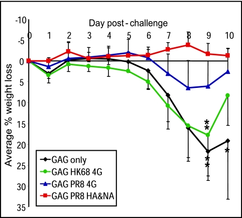
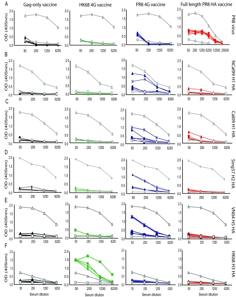
Although highly effective in the general population when well matched to circulating influenza virus strains, current influenza vaccines are limited in their utility due to the narrow breadth of protection they provide. The strain specificity of vaccines presently in use mirrors the exquisite specificity of the neutralizing antibodies that they induce, that is, antibodies which bind to the highly variable globular head domain of hemagglutinin (HA). Herein, we describe the construction of a novel immunogen comprising the conserved influenza HA stalk domain and lacking the globular head. Vaccination of mice with this headless HA construct elicited immune sera with broader reactivity than those obtained from mice immunized with a full-length HA. Furthermore, the headless HA vaccine provided full protection against death and partial protection against disease following lethal viral challenge. Our results suggest that the response induced by headless HA vaccines is sufficiently potent to warrant their further development toward a universal influenza virus vaccine.


Figures

FIG 1
FIG 1
Schematic of headless HA constructs. (A) Schematic of the linear structure of the full-length influenza virus HA protein (top) and a generalized headless HA protein (bottom). Linker peptides tested in the context of the PR8 and HK68 HA sequences are shown. Inserted amino acids are shown in bold red type, while amino acids present in the native HA sequence are in black. (B) Schematic of the folded structures of the full-length and headless HAs of PR8 virus (left panel) and HK68 virus (right panel). In both cases, headless HAs carrying the 4G linker bridge are depicted. The HA1 subunit is blue, and the HA2 subunit is green. Cysteines 52 and 277 are shown in yellow, and 4G linker sequences are shown in red. The full-length HA structures were downloaded from the Protein Database (PDB): PR8 HA (PDB ID 1RVX) and HK68 HA (PDB ID 1MQN). Schematics of headless HAs were generated using the full-length HA coordinates as a starting point, and 4G loops were manually docked into the headless HA carbon to close the discontinuous alpha-carbon amino acid chain. Final images were generated with PyMol (Delano Scientific).
FIG 2
FIG 2
Expression of headless HA constructs in transiently transfected cells. Headless HA constructs were expressed in 293T cells by plasmid transfection in the absence of exogenous trypsin. At 24 h posttransfection, whole-cell lysates were prepared and subjected to sodium dodecyl sulfate-polyacrylamide gel electrophoresis (SDS-PAGE) followed by Western blotting. HA proteins were detected using the polyclonal antiserum 3951 (for PR8) or the MAb 12D1 (for HK68). Molecular mass markers in kilodaltons are shown to the left of each blot, and transfected constructs are identified above the appropriate lane. Mock, untransfected cells; full, full-length HA protein. For the headless HA constructs, the amino acid sequence bridging the N- and C-terminal strands of HA1 is shown. Boldface letters indicate inserted amino acids, while lightface letters represent residues present in the wild-type HA. In the region of the Cys 52 to Cys 277 disulfide bond, the wild-type sequences are as follows: for PR8, K50L51C52…C277N278T279K280; for HK68, K50I51C52… C277I278S279E280. (A) PR8-based constructs; (B) HK68-based constructs. In panel B, the molecular weight of the full-length HK68 HA0 protein is indicated by an arrow.
FIG 3
FIG 3
Detection of headless HA proteins on the surfaces of transfected cells. Full-length and headless HA constructs were expressed in 293T cells by plasmid transfection. At 24 h posttransfection, cells were trypsinized and HA proteins on the cell surface were stained using polyclonal antiserum 3951 (for PR8) or MAb 12D1 (for HK68) before analysis by flow cytometry. (A) Mock-transfected cells stained with immune serum 3951 (gray) are compared to cells transfected with pDZ PR8 HA (red) or cells transfected with pCAGGS PR8 2G, 4G, or PG headless HA constructs (blue). (B) Mock-transfected cells stained with MAb 12D1 (gray) are compared to cells transfected with pCAGGS HK68 HA (orange) or cells transfected with pCAGGS HK68 2G, 4G, or PG headless HA constructs (green).
FIG 4
FIG 4
Incorporation of headless HA proteins into VLPs. The HA content of VLPs generated by cotransfection of HA constructs with pGag-EGFP was assessed by Western blotting. PR8-based VLPs were probed with polyclonal antiserum 3951, and HK68-based proteins were detected with MAb 12D1. Bands are identified to the right of each blot. Note that VLPs were produced in the presence of exogenous trypsin, resulting in the cleavage of HA0 to produce HA1 (not visualized here) and HA2. Each lane shows VLPs harvested from the equivalent of one 10-cm dish.
FIG 5
FIG 5
Vaccination of mice with headless HA constructs provides protection from death. The average body weight loss in each group of vaccinated mice following challenge with PR8 virus is shown. Error bars represent standard deviations. A star indicates the death of a mouse.
FIG 6
FIG 6
Antisera from mice vaccinated with the PR8 4G headless HA show broad cross-reactivity by ELISA. The vaccine groups from which sera are derived are identified at the top of each column, and the ELISA substrate used is indicated to the right of each row. The colors used are consistent throughout the figure, with sera from Gag-only-vaccinated mice indicated in black, HK68 4G-vaccinated mice indicated in green, PR8 4G-vaccinated mice indicated in blue, and full-length-PR8-HA-vaccinated mice indicated in red. Each mouse is represented by a unique symbol that is the same in each panel. A rabbit antiserum raised against whole PR8 virus is shown in gray with open triangles, and a serum sample taken from a naive mouse is shown in gray with open squares. The reactivities of mouse sera to whole PR8 virus (A), purified recombinant A/New Caledonia/20/1999 HA protein (B), purified recombinant A/California/04/2009 HA (C), purified recombinant A/Singapore/1/1957 HA (D), purified recombinant A/Viet Nam/ 1203/2004 HA (E), and purified recombinant A/Hong Kong/ 1/1968 HA (F) are shown.

Similar articles

Cited by

References

    1. Fox J. P., Cooney M. K., Hall C. E., Foy H. M. 1982. Influenza virus infections in Seattle families, 1975-1979. II. Pattern of infection in invaded households and relation of age and prior antibody to occurrence of infection and related illness. Am. J. Epidemiol. 116:228–242 - PubMed
    1. Glezen W. P., Couch R. B. 1978. Interpandemic influenza in the Houston area, 1974-1976. N. Engl. J. Med. 298:587–592 - PubMed
    1. Belshe R. B. 2007. Translational research on vaccines: influenza as an example. Clin. Pharmacol. Ther. 82:745–749 - PubMed
    1. Thompson W. W., Shay D. K., Weintraub E., Brammer L., Cox N., Anderson L. J., Fukuda K. 2003. Mortality associated with influenza and respiratory syncytial virus in the United States. JAMA 289:179–186 - PubMed
    1. Johnson N. P., Mueller J. 2002. Updating the accounts: global mortality of the 1918-1920 “Spanish” influenza pandemic. Bull. Hist. Med. 76:105–115 - PubMed

LinkOut - more resources