Replication timing aberrations and aneuploidy in peripheral blood lymphocytes of breast cancer patients
- PMID: 20689761
- PMCID: PMC2915411
- DOI: 10.1593/neo.10568
Replication timing aberrations and aneuploidy in peripheral blood lymphocytes of breast cancer patients
Abstract
Background: Peripheral blood lymphocytes of patients with hematological malignancies or solid tumors, such as renal cell carcinoma or prostate cancer, display epigenetic aberrations (loss of synchronous replication of allelic counterparts) and genetic changes (aneuploidy) characteristic of the cancerous phenotype. This study sought to determine whether such alterations could differentiate breast cancer patients from cancer-free subjects.
Methods: The HER2 locus-an oncogene assigned to chromosome 17 whose amplification is associated with breast cancer (BCA)-and the pericentromeric satellite sequence of chromosome 17 (CEN17) were used for replication timing assessments. Aneuploidy was monitored by enumerating the copy numbers of chromosome 17. Replication timing and aneuploidy were detected cytogenetically using fluorescence in situ hybridization technology applied to phytohemagglutinin-stimulated lymphocytes of 20 women with BCA and 10 control subjects.
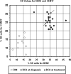
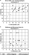
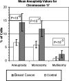
Results: We showed that both the HER2 and CEN17 loci in the stimulated BCA lymphocytes altered their characteristic pattern of synchronous replication and exhibited asynchronicity. In addition, there was an increase in chromosome 17 aneuploidy. The frequency of cells displaying asynchronous replication in the patients' samples was significantly higher (P < 10(-12) for HER2 and P < 10(-6) for CEN17) than the corresponding values in the control samples. Similarly, aneuploidy in patients' cells was significantly higher (P < 10(-9)) than that in the controls.
Conclusions: The HER2 and CEN17 aberrant replication differentiated clearly between BCA patients and control subjects. Thus, monitoring the replication of these genes offers potential blood markers for the detection and monitoring of breast cancer.
Figures


Similar articles
-
The aberrant asynchronous replication - characterizing lymphocytes of cancer patients - is erased following stem cell transplantation.BMC Cancer. 2010 May 24;10:230. doi: 10.1186/1471-2407-10-230. BMC Cancer. 2010. PMID: 20497575 Free PMC article.
-
Asynchronous DNA replication and aneuploidy in lymphocytes of hepatocellular carcinoma patients.Cancer Genet. 2012 Dec;205(12):636-43. doi: 10.1016/j.cancergen.2012.10.006. Epub 2012 Nov 23. Cancer Genet. 2012. PMID: 23182962
-
Altered mode of allelic replication accompanied by aneuploidy in peripheral blood lymphocytes of prostate cancer patients.Int J Cancer. 2004 Aug 10;111(1):60-6. doi: 10.1002/ijc.20237. Int J Cancer. 2004. PMID: 15185343
-
DNA amplifications in breast cancer: genotypic-phenotypic correlations.Future Oncol. 2010 Jun;6(6):967-84. doi: 10.2217/fon.10.56. Future Oncol. 2010. PMID: 20528234 Review.
-
[Diagnosis of HER2 gene amplification in breast carcinoma].Pathol Biol (Paris). 2008 Sep;56(6):375-9. doi: 10.1016/j.patbio.2008.03.009. Epub 2008 May 5. Pathol Biol (Paris). 2008. PMID: 18456424 Review. French.
Cited by
-
Chromosome mis-segregation and cytokinesis failure in trisomic human cells.Elife. 2015 May 5;4:e05068. doi: 10.7554/eLife.05068. Elife. 2015. PMID: 25942454 Free PMC article.
-
Chromosomal Rearrangements and Altered Nuclear Organization: Recent Mechanistic Models in Cancer.Cancers (Basel). 2021 Nov 22;13(22):5860. doi: 10.3390/cancers13225860. Cancers (Basel). 2021. PMID: 34831011 Free PMC article. Review.
-
Perturbations in the Replication Program Contribute to Genomic Instability in Cancer.Int J Mol Sci. 2017 May 25;18(6):1138. doi: 10.3390/ijms18061138. Int J Mol Sci. 2017. PMID: 28587102 Free PMC article. Review.
-
The interconnectedness of cancer cell signaling.Neoplasia. 2011 Dec;13(12):1183-93. doi: 10.1593/neo.111746. Neoplasia. 2011. PMID: 22241964 Free PMC article.
-
DNA replication timing, genome stability and cancer: late and/or delayed DNA replication timing is associated with increased genomic instability.Semin Cancer Biol. 2013 Apr;23(2):80-9. doi: 10.1016/j.semcancer.2013.01.001. Epub 2013 Jan 14. Semin Cancer Biol. 2013. PMID: 23327985 Free PMC article. Review.
References
-
- Gilbert DM. Replication timing and transcriptional control: beyond cause and effect. Curr Opin Cell Biol. 2002;14:377–383. - PubMed
-
- McNairn AJ, Gilbert DM. Epigenomic replication: linking epigenetics to DNA replication. Bioessays. 2003;25:647–656. - PubMed
-
- Woodfine K, Fiegler H, Beare DM, Collins JE, McCann OT, Young BD, Debernardi S, Mott R, Dunham I, Carter NP. Replication timing of the human genome. Hum Mol Genet. 2004;13:191–202. - PubMed
-
- Goren E, Cedar H. Replication by the clock. Nat Rev Mol Cell Biol. 2003;4:25–32. - PubMed
MeSH terms
LinkOut - more resources
Full Text Sources
Other Literature Sources
Medical
Research Materials
Miscellaneous
