

Electroretinographic findings in transplant chorioretinopathy
- PMID: 20689794
- PMCID: PMC2915864
- DOI: 10.2147/opth.s12057
Electroretinographic findings in transplant chorioretinopathy
Abstract
Aim: Transplant chorioretinopathy is a rare complication following solid organ or bone marrow transplantation and can result in severe vision loss. This series presents electroretinogram (ERG) results in patients with this condition.
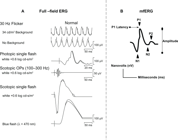
Methods: Patients who presented with bilateral vision loss following bone marrow or solid organ transplantation were identified. A complete ophthalmologic examination, fundus photography, and fluorescein angiography (FA) were performed. Full-field ERG was obtained in all patients and a multifocal ERG (mfERG) was obtained in two patients.
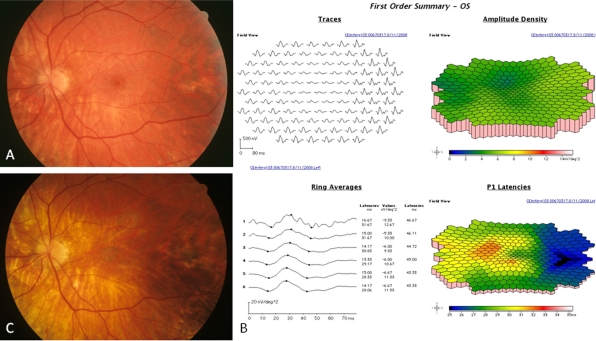
Results: Four patients were identified. All patients had bilateral vision loss and displayed a characteristic pattern of mottled hyperfluorescence on FA. Three patients developed progressive vision loss ranging from 20/60 to hand motions whereas one retained 20/40 vision. All patients exhibited moderate to severe cone dysfunction, while the degree of rod abnormalities was varied. Two patients with severe cone dysfunction showed mild clinical changes initially, but later developed progressive vision loss and chorioretinal atrophy.
Conclusion: Transplant chorioretinopathy patients undergoing ERG testing show cone dysfunction with a variable degree of rod dysfunction. ERG abnormalities preceded the visual acuity and clinical changes in two patients, suggesting that ERG may be a helpful predictor of the clinical course in this rare disease.
Keywords: ERG; chorioretinopathy; electroretinogram; mfERG; transplant.
Figures


Similar articles
-
Retinal dysfunction in a presymptomatic patient with Huntington's disease.Doc Ophthalmol. 2018 Jun;136(3):213-221. doi: 10.1007/s10633-018-9632-3. Epub 2018 Apr 24. Doc Ophthalmol. 2018. PMID: 29691705
-
Electroretinographic findings in patients with Stargardt disease and fundus flavimaculatus.Retina. 2004 Dec;24(6):920-8. doi: 10.1097/00006982-200412000-00013. Retina. 2004. PMID: 15579991
-
Late onset cone dystrophy.Doc Ophthalmol. 2010 Jun;120(3):215-8. doi: 10.1007/s10633-010-9214-5. Epub 2010 Jan 13. Doc Ophthalmol. 2010. PMID: 20069340
-
Ophthalmologic findings in long-chain 3-hydroxyacyl-CoA dehydrogenase deficiency caused by the G1528C mutation: a new type of hereditary metabolic chorioretinopathy.Ophthalmology. 1998 May;105(5):810-24. doi: 10.1016/S0161-6420(98)95019-9. Ophthalmology. 1998. PMID: 9593380
-
Electroretinographic monitoring in birdshot chorioretinopathy.Am J Ophthalmol. 2005 Jul;140(1):52-64. doi: 10.1016/j.ajo.2005.01.053. Am J Ophthalmol. 2005. PMID: 16038651
References
-
- Gass JDM, Slamvotis TL, Full DG, et al. Posterior chorioretinopathy and retinal detachment after organ transplantation. Arch Ophthalmol. 1992;110(12):1717–1722. - PubMed
-
- Gass JDM. Stereoscopic Atlas of Macular Diseases: Diagnosis and Treatment. 4th ed. St. Louis, MO: Mosby; 1997.
-
- Fawzi AA, Cunningham ET., Jr Central serous chorioretinopathy after bone marrow transplantation. Am J Ophthalmol. 2001;131(6):804–805. - PubMed
-
- Fawzi AA, Holland GN, Kreiger AE, et al. Central serous chorioretinopathy after solid organ transplantation. Ophthalmology. 2006;113(5):805–813. - PubMed
-
- Friberg TR, Eller AW. Serous retinal detachment resembling central serous chorioretinopathy following organ transplantation. Graefes Arch Clin Exp Ophthalmol. 1990;228(4):305–309. - PubMed
Publication types
Grants and funding
LinkOut - more resources
Full Text Sources
