

Monozygotic twins with polypoidal choroidal vasuculopathy
- PMID: 20689796
- PMCID: PMC2915866
- DOI: 10.2147/opth.s11003
Monozygotic twins with polypoidal choroidal vasuculopathy
Abstract
Purpose: To present the first findings in the set of monozygotic twins with polypoidal choroidopathy (PCV).
Methods: Sixty two-year old monozygotic twin sisters were studied. The concordances and discordances of the clinical features of the twins were determined. Genomic DNA was extracted and genotyped for three established PCV risk-associated single nucleotide polymorphisms, viz CFH I62V, CFH Y402H, and ARMS A69S.
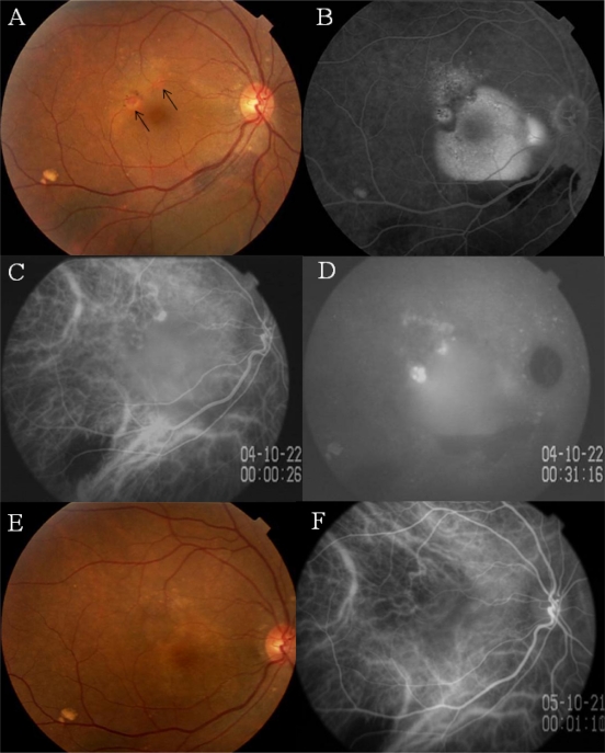
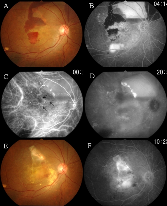
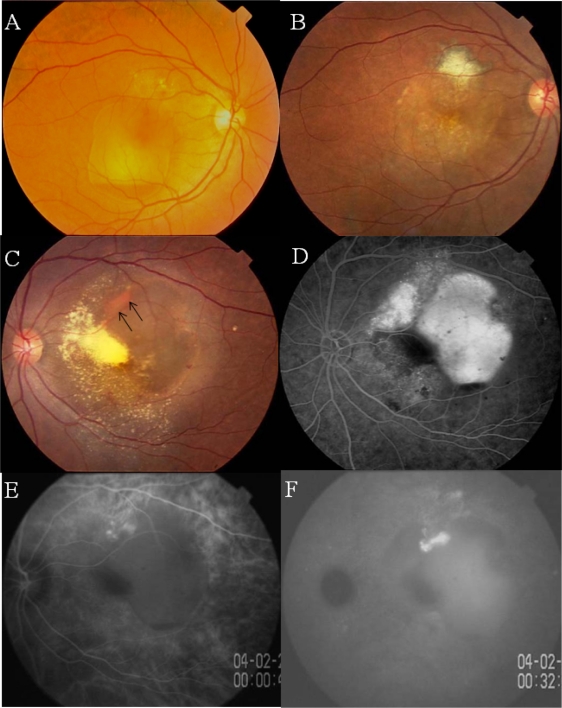
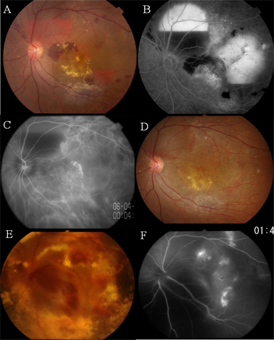
Results: Both patients had hemorrhagic pigment epithelial detachments with orange lesions beneath the retinal pigment epithelium. Indocyanine green angiography showed pathognomonic choroidal vascular networks with polypoidal structures uniocularly in one twin and binocularly in the other twin. Both twins were treated with photodynamic therapy, retinal photocoagulation, and anti-vascular endothelial growth factor therapy, but both showed limited response to all the treatments, with recurrent exudative lesions with enlarged vascular network, and poor visual outcome. Genetic analyses showed that both sisters had homozygous risk alleles for ARMS2 A69S, and one risk allele each of CFH I62V and CFH Y402H.
Conclusions: We present the first findings in a set of monozygotic twins with typical PCV under long-term observation. The concordances in disease progression and response to treatment between the twins indicate that these genetic factors most likely played important roles in determining the clinical manifestations.
Keywords: ARMS2; CFH; PCV; monozygotic twins; polypoidal choroidal vasculopathy.
Figures
References
-
- Yannuzzi LA, Sorenson J, Spaide RF, et al. Idiopathic polypoidal choroidopathy (IPCV) Retina. 1990;10:1–8. - PubMed
-
- Spaide RF, Yannuzzi LA, Slakter JS, et al. Indocyanine green videoangiography of idiopathic polypoidal choroidal vasculopathy. Retina. 1995;15:100–110. - PubMed
-
- Laude A, Cackett PD, Vithana EN, et al. Polypoidal choroidal vasculopathy and neovascular age-related macular degeneration: same or different disease? Prog Retin Eye Res. 2010;29:19–29. - PubMed
-
- Mori K, Horie-Inoue K, Gehbach PL, et al. Phenotype and genotype characteristics of age-related macular degeneration in a Japanese population. Ophthalmology. 2010, Epub ahead of print. - PubMed
-
- Kikuchi M, Nakamura M, Ishikawa K, et al. Elevated C-reactive protein levels in patients with polypoidal choroidal vasculopathy and patients with neovascular age-related macular degeneration. Ophthalmology. 2007;114:1722–1727. - PubMed
Publication types
LinkOut - more resources
Full Text Sources
Other Literature Sources
Miscellaneous
