. 2010 Aug 3;8(8):e1000442.
doi: 10.1371/journal.pbio.1000442.

Rac1-dependent collective cell migration is required for specification of the anterior-posterior body axis of the mouse

Affiliations

Rac1-dependent collective cell migration is required for specification of the anterior-posterior body axis of the mouse

Isabelle Migeotte et al. PLoS Biol. .

Abstract

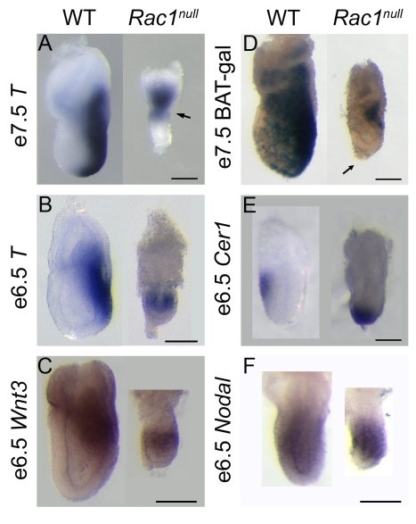
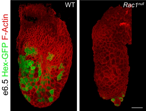
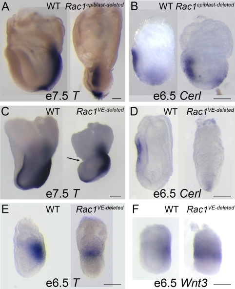
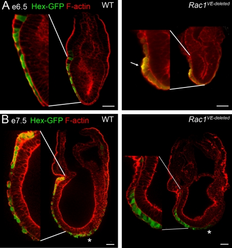
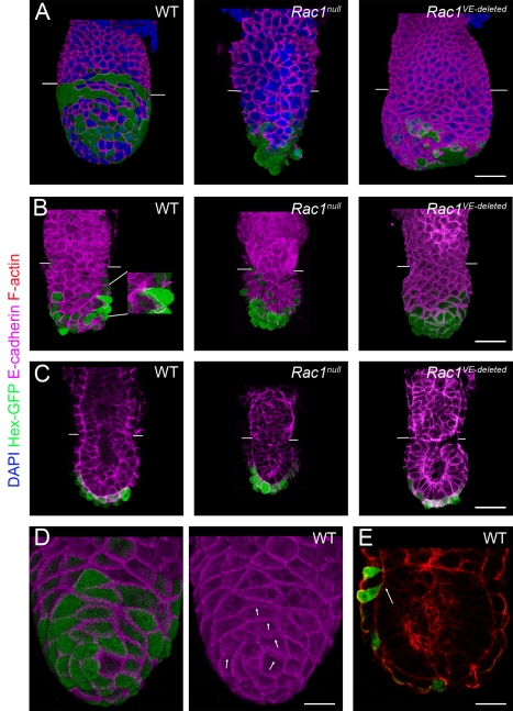
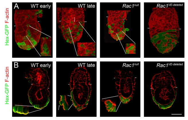
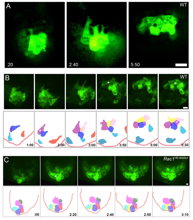
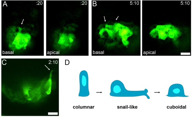
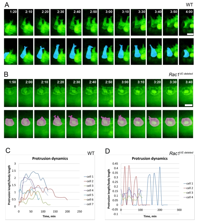
Cell migration and cell rearrangements are critical for establishment of the body plan of vertebrate embryos. The first step in organization of the body plan of the mouse embryo, specification of the anterior-posterior body axis, depends on migration of the anterior visceral endoderm from the distal tip of the embryo to a more proximal region overlying the future head. The anterior visceral endoderm (AVE) is a cluster of extra-embryonic cells that secretes inhibitors of the Wnt and Nodal pathways to inhibit posterior development. Because Rac proteins are crucial regulators of cell migration and mouse Rac1 mutants die early in development, we tested whether Rac1 plays a role in AVE migration. Here we show that Rac1 mutant embryos fail to specify an anterior-posterior axis and, instead, express posterior markers in a ring around the embryonic circumference. Cells that express the molecular markers of the AVE are properly specified in Rac1 mutants but remain at the distal tip of the embryo at the time when migration should take place. Using tissue specific deletions, we show that Rac1 acts autonomously within the visceral endoderm to promote cell migration. High-resolution imaging shows that the leading wild-type AVE cells extend long lamellar protrusions that span several cell diameters and are polarized in the direction of cell movement. These projections are tipped by filopodia-like structures that appear to sample the environment. Wild-type AVE cells display hallmarks of collective cell migration: they retain tight and adherens junctions as they migrate and exchange neighbors within the plane of the visceral endoderm epithelium. Analysis of mutant embryos shows that Rac1 is not required for intercellular signaling, survival, proliferation, or adhesion in the visceral endoderm but is necessary for the ability of visceral endoderm cells to extend projections, change shape, and exchange neighbors. The data show that Rac1-mediated epithelial migration of the AVE is a crucial step in the establishment of the mammalian body plan and suggest that Rac1 is essential for collective migration in mammalian tissues.


Conflict of interest statement

The authors have declared that no competing interests exist.

Figures

Figure 1
Figure 1. Rac1 null embryos fail to specify an AP axis.
Expression markers of the primitive streak and AVE. (A) Wild-type e7.5 embryos expressed Brachyury (T) in the primitive streak, on the posterior side of the embryo. E7.5 Rac1 null embryos displayed a constriction at the embryonic/extra-embryonic boundary (arrow). They expressed T in a ring at the embryonic/extra-embryonic border (6/8) or only in the extra-embryonic region (not shown; 2/8). In 2 out of 8 embryos, T expression was slightly polarized to one side. (B) At e6.5, T was expressed in a spot (2/17) or ring (9/17) at the embryonic/extra-embryonic border of Rac1 null embryos. No expression was detected in 6/17 mutants, presumably due to developmental delay. (C) Wnt3 was expressed around the circumference of Rac1 null embryos at e6.5 (7/17; no staining in 10/17). (D) In 2/2 mutant embryos, the Wnt reporter BAT-gal was expressed in a slightly polarized fashion at the embryonic/extra-embryonic junction at e7.5. Note that in those embryos where the primitive streak was asymmetric, a cluster of AVE cells that failed to migrate (arrow) was slightly displaced from the embryo tip opposite to the position of streak marker expression, revealing a slight residual asymmetry. (E) Cer1, marking the AVE, is expressed distally at e6.5 in Rac1 null embryos. (F) Nodal, which is required for specification of the AVE, was expressed at e6.5 in a proximal-to-distal gradient in Rac1 null embryos. WT, wild-type. Scale bars = 100 µm in A, B, C, D, and F, and 50 µm in E. Anterior is to the left in all panels.
Figure 2
Figure 2. Hex-expressing AVE cells fail to migrate in Rac1 null embryos.
3D reconstruction of whole-mount confocal Z-stacks of e6.5 embryos stained with phalloidin to visualize F-actin (red). Expression of the Hex-GFP transgene (green) marks AVE cells at the end of migration. Hex-expressing cells reside at the embryonic/extra-embryonic boundary of wild-type embryos at this stage. In contrast, Hex-expressing cells are found in a cluster at the distal tip of the Rac1 mutant. Hex-GFP was detected with anti-GFP antibody in wild-type and native GFP in the mutant. Scale bar = 50 µm.
Figure 3
Figure 3. Rac1 acts autonomously to promote AVE migration.
In situ hybridization with markers of the AVE and the primitive streak; lateral views of embryos are shown. (A) T was expressed normally in e7.5 Rac1 epiblast-deleted embryos, although the shape of the primitive streak was disrupted in the mutants. The T-positive primitive streak formed a bulge that filled most of the amniotic cavity, due to disruption of the behavior of the embryonic mesoderm (; Migeotte and Anderson, in preparation). (B) Cer1 was expressed at the normal position in Rac1 epiblast-deleted embryos. (C) In embryos where Rac1 was deleted from the VE using Ttr-Cre, there was a strong constriction at the embryonic/extra-embryonic boundary (arrow) and abnormal headfolds. Axis specification, as marked by T expression, appeared normal at e7.5. (D) At e6.5, Cer1 was expressed close to the distal tip of the embryo in the majority of Rac1 VE-deleted embryos (4/7) and had reached the extra-embryonic boundary in 3/7. (E) T was expressed in a ring in 3/10 Rac1 VE-deleted embryos at e6.5 and in a spot on the posterior side of the embryo in 7/10 embryos. (F) Wnt3 was expressed in a ring in 2/2 Rac1 VE-deleted embryos at e6.5. Scale bars = 100 µm.
Figure 4
Figure 4. Epithelial organization of Rac1 VE-deleted embryos.
Individual confocal sections of whole-mount embryos stained for F-actin (red). AVE cells were marked with the Hex-GFP transgene (green, staining with anti-GFP antibody in A and native GFP in B). After AVE migration is complete, at e6.5 (A) and e7.5 (B), AVE cells formed a single-layer epithelium and were squamous in wild-type embryos, with the exception of the most proximal cells, which were cuboidal. At e7.5, definitive endoderm cells expressing Hex-GFP (marked by an *) were present at the distal end of the primitive streak. In VE-deleted embryos, AVE cells had failed to reach the embryonic/extra-embryonic boundary. The Hex-GFP-expressing cells retained the columnar shape characteristic of migrating AVE cells and displayed a strong apical actin at e6.5 (arrow in A). Scale bars = 50 µm. Insets are 2×.
Figure 5
Figure 5. E-cadherin during migration.
(A, B) 3D reconstructions showing expression of Hex-GFP (green, staining with anti-GFP antibody) and E-cadherin (magenta). See Figure S2 for the separated E-cadherin channel. (A) Embryos at the time when migration should be nearly completed. Some wild-type AVE cells have reached the extra-embryonic boundary (marked by white lines in all embryos), while they remain distal in stage-matched Rac1 null and Rac1 VE-deleted embryos (dissected at e6.25). (B) e5.5–e5.75 embryos during the time of AVE migration. Wild-type and mutant Hex-GFP cells had comparable levels of E-cadherin. Migrating wild-type embryonic VE cells had diverse shapes, and some cells with long projections could be observed (inset, 2×). In Rac1 null embryos, cells were more regular and rounder. Rac1 VE-deleted embryos had a variable phenotype at e5.75. Some mutants were indistinguishable from wild-type (5/14), some had AVE cells that migrated from distal to proximal but failed to spread laterally (5/14), and some (shown) displayed a partial distal to proximal migration (4/14). (C) Individual sections from Z-stacks of e5.5–e5.75 embryos, stained for E-cadherin. Wild-type AVE cells retained adherens junctions during migration. E5.5 Rac1 null and Rac1 VE-deleted embryos displayed a single-layered epithelium with normal-appearing adherens junctions. (D) 3D reconstructions of Z-stack of a wild-type e5.5 embryo. The lateral membrane of some cells was at an oblique angle to the basement membrane, causing the E-cadherin staining to appear fuzzy (arrows). (E) Individual section from a Z-stack of a wild-type e5.75 embryo stained with phalloidin to visualize F-actin (red). AVE cells on the proximal anterior surface of the embryo are tilted in the apical/basal plane, so that their basal surfaces (arrow) are more anterior than their apical surface. Scale bars = 50 µm in A to C, and 25 µm in D and E.
Figure 6
Figure 6. F-actin during migration.
(A) Anterior views of 3D reconstructions of Z-stacks of e5.5–e5.75 embryos expressing Hex-GFP (green, staining with anti-GFP antibody), stained for F-actin (red). The embryonic/extra-embryonic boundary is marked by white lines in all embryos. The early e5.5 wild-type embryo was cultured for 1 h prior to fixation; these conditions favored the preservation of long protrusions. Wild-type embryonic VE cells had diverse shapes during migration (inset, 1.5×), while in Rac1 null (inset, 1.5×) and Rac1 VE-deleted embryos, cell shapes were more regular and rounder. Wild-type VE cells formed vertices of up to 7 cells (arrow, inset, 1.5×). (B) Individual frontal sections from Z-stacks of e5.5–e5.75 embryos expressing Hex-GFP (detected with anti-GFP antibody). F-actin staining was weaker in the apical surface of migrating cells (arrow), while in wild-type pre-migration cells and in mutant cells apical F-actin staining was retained (insets, 2×). Scale bars = 50 µm.
Figure 7
Figure 7. AVE cells send long lamellipodia that are spatially and temporally coordinated and depend on Rac1.
Hex-GFP expressing embryos were dissected at e5.5 and cultured for up to 18 h. Proximal is up in all panels. (A) Individual confocal sections extracted from an 18 h live imaging experiment (Video S1). Four Hex-positive cells simultaneously sent long, parallel projections toward the extra-embryonic region. By 5 h 50 min, the cells reached the boundary of the extra-embryonic region, and the protrusive activity stopped. Images were aligned relative to the distal border of the embryo. (B) Hex-GFP-positive cells migrated from distal to proximal in a coordinated fashion, and then spread laterally on the anterior surface of the embryo. In the lower panel, cells from maximal projection images from stacks of confocal sections (stills from Video S1) were colored to follow individual cell movements and changes of neighbors. Asterisks show the intercalations of previously hidden cells. (C) In Rac1 VE-deleted embryos there was no cell migration, and no exchange of neighbors (lower panel). Asterisk shows the intercalation of a previously hidden cell. In the lower panel, cells from maximal projection images from stacks of confocal sections (stills from Video S11) were colored. Scale bars = 30 µm.
Figure 8
Figure 8. AVE cells insert basal projections between neighboring cells to move forward.
(A) Individual confocal sections of a migrating snail-like AVE cell (dark blue cell from Video S1). The basal section shows a long lamella with two projections at its end (arrow); the projection is not visible apically. The distance between the apical and basal optical sections is 8 µm. (B) Individual confocal sections of an AVE cell (yellow cell from Video S1) that has arrived at the embryonic/extra-embryonic border. The cell shape became cuboidal. The basal surface was marked by retracting protrusions (arrow) that are not visible in the apical plane. The distance between the apical and basal optical sections is 14 µm. Scale bar = 30 µm. (C) Individual confocal section of an AVE cell from Video S1, oriented to show the apical-basal profile of the cell. The long projection is on the basal side. (D) Schematic of the shape changes of the cells with long lamellar projections. At e5.5, AVE cells are columnar. During migration, cells acquire a snail-like morphology with a round cell body and a highly elongated lamella ended by two lateral horn-like protrusions. At the end of migration, the cells become cuboidal.
Figure 9
Figure 9. The long stable protrusions of AVE cells are Rac1-dependent.
(A, B) Individual confocal sections were aligned against the distal border of the embryo (A) or against the cell body (B), and painted to highlight cell shape in the lower row. (A) The wild-type cell displayed is the purple cell in Figure 7B. The protrusive activity persisted for 2 h. The long protrusions had a stable direction for about 30 min (2 h 10 min–2 h 40 min), then retracted partially and extended at a slightly different angle (3 h 10 min–3 h 40 min) (Videos S1 and S6). (B) The Rac1 mutant cell displayed is the pink cell in Figure 7C. The cell was non-polarized and rounded. The small protrusions of mutant cells were unstable (Videos S11 and S13). Scale bar = 30 µm. (C, D) Quantification of protrusion length and stability. (C) In wild-type embryos, the protrusions lasted for at least 1 h and their length reached up to 2.5 times the diameter of the cell body (defined as the circular region of the cell surrounding the nucleus). 7 cells from 5 experiments were analyzed. (D) In Rac1 VE-deleted embryos protrusions (4 cells from 2 experiments) were short (less than half the cell body diameter) and transient (∼10 min).

Similar articles

Cited by

References

    1. Thomas P. Q, Brown A, Beddington R. S. Hex: a homeobox gene revealing peri-implantation asymmetry in the mouse embryo and an early transient marker of endothelial cell precursors. Development. 1998;125:85–94. - PubMed
    1. Srinivas S, Rodriguez T, Clements M, Smith J. C, Beddington R. S. P. Active cell migration drives the unilateral movements of the anterior visceral endoderm. Development. 2004;131:1157–1164. - PubMed
    1. Kimura-Yoshida C, Nakano H, Okamura D, Nakao K, Yonemura S, et al. Canonical Wnt signaling and its antagonist regulate anterior-posterior axis polarization by guiding cell migration in mouse visceral endoderm. Developmental Cell. 2005;9:639–650. - PubMed
    1. Srinivas S. The anterior visceral endoderm-turning heads. Genesis. 2006;44:565–572. - PubMed
    1. Salgueiro A. M, Filipe M, Belo J. A. N-acetylgalactosamine 4-sulfate 6-O-sulfotransferase expression during early mouse embryonic development. Int J Dev Biol. 2006;50:705–708. - PubMed

Publication types

MeSH terms