. 2010 Aug 3;5(8):e11929.
doi: 10.1371/journal.pone.0011929.

A critical tryptophan and Ca2+ in activation and catalysis of TPPI, the enzyme deficient in classic late-infantile neuronal ceroid lipofuscinosis

Affiliations

A critical tryptophan and Ca2+ in activation and catalysis of TPPI, the enzyme deficient in classic late-infantile neuronal ceroid lipofuscinosis

Salomon Kuizon et al. PLoS One. .

Abstract

Background: Tripeptidyl aminopeptidase I (TPPI) is a crucial lysosomal enzyme that is deficient in the fatal neurodegenerative disorder called classic late-infantile neuronal ceroid lipofuscinosis (LINCL). It is involved in the catabolism of proteins in the lysosomes. Recent X-ray crystallographic studies have provided insights into the structural/functional aspects of TPPI catalysis, and indicated presence of an octahedrally coordinated Ca(2+).

Methodology: Purified precursor and mature TPPI were used to study inhibition by NBS and EDTA using biochemical and immunological approaches. Site-directed mutagenesis with confocal imaging technique identified a critical W residue in TPPI activity, and the processing of precursor into mature enzyme.

Principal findings: NBS is a potent inhibitor of the purified TPPI. In mammalian TPPI, W542 is critical for tripeptidyl peptidase activity as well as autocatalysis. Transfection studies have indicated that mutants of the TPPI that harbor residues other than W at position 542 have delayed processing, and are retained in the ER rather than transported to lysosomes. EDTA inhibits the autocatalytic processing of the precursor TPPI.

Conclusions/significance: We propose that W542 and Ca(2+) are critical for maintaining the proper tertiary structure of the precursor proprotein as well as the mature TPPI. Additionally, Ca(2+) is necessary for the autocatalytic processing of the precursor protein into the mature TPPI. We have identified NBS as a potent TPPI inhibitor, which led in delineating a critical role for W542 residue. Studies with such compounds will prove valuable in identifying the critical residues in the TPPI catalysis and its structure-function analysis.


Conflict of interest statement

Competing Interests: The authors have declared that no competing interests exist.

Figures

Figure 1
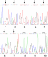
Figure 1. Site-directed mutagenesis of W residues in TPPI and DNA sequencing.
W residues were numbered 1–7 starting from the amino-terminus. The corresponding amino acid positions in the mature TPPI are 1, W290; 2, W307; 3, W366; 4, W460; 5, W470; 6, W542; and 7, W548. Arrows indicate the nucleotide change from g→t. Sequence numbers 8–10 are W542D, H, and K mutants, respectively. The bar represents the codon replaced to change the W residue.
Figure 2
Figure 2. Specific activity of TPPI for various W mutants relative to mature TPPI.
Freshly plated CHO cells at 70% confluency were transiently transfected with mutant plasmids using FugeneHD reagent. After 48 h, cells were pelleted by centrifugation at 800× g for 5 min, followed by homogenization in lysis buffer comprising 50 mM ammonium formate, pH 3.5, containing 0.15M NaCl and 0.1% Triton X-100. TPPI activity was measured as described in Experimental Procedures and related to the band intensity of mature TPPI by Western blot analysis.
Figure 3
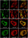
Figure 3. Localization of wt and mutant TPPI in CHO cells.
Freshly plated CHO cells at 70% confluency were transiently transfected with mutant plasmids using FugeneHD reagent. After 24 h, cells were immunostained for TPPI (8C4, monoclonal) and calreticulin (rabbit polyclonal). Secondary antibodies labeled with Alexa Fluor 488 (green fluorescence visualizing TPPI) and Alexa Fluor 555 (red fluorescence visualizing calreticulin) were used. Upon merging, the yellow color indicates overlap of expressed proteins. Digital images were taken at a magnification of ×1000.
Figure 4
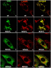
Figure 4. Localization of wt and mutant TPPI in CHO cells.
Freshly plated CHO cells at 70% confluency were transiently transfected with mutant plasmids using FugeneHD reagent. After 24 h, cells were immunostained for TPPI (RAS307, rabbit polyclonal) and LAMP (mouse monoclonal). Secondary antibodies labeled with Alexa Fluor 488 (green fluorescence visualizing TPPI) and Alexa Fluor 555 (red fluorescence visualizing LAMP) were used. Upon merging, the yellow color indicates overlap of expressed proteins. Digital images were taken at a magnification of ×1000.
Figure 5
Figure 5. Autocatalytic processing of pro-TPPI.
Western blot analysis of the expression and processing of wt and various mutant TPPI cDNAs in transiently transfected CHO cells. CHO cells at confluency were transiently transfected with various plasmids using the FugeneHD reagent. After 48 h, cells were homogenized in sample buffer, and total cellular proteins (25 µg) were resolved on 10% SDS-PAGE. After transferring onto nitrocellulose membrane, TPPI was visualized by chemiluminesce using mouse monoclonal antibody (8C4). Lane 1, wt TPPI; lane 2, mutant W290L; lane 3, mutant W307L; lane 4, mutant W366L; lane 5, mutant W460L; lane 6, mutant W470L; lane 7, mutant W542L; lane 8, mutant W548L; lane 9, mutant W542D; lane 10, mutant W542H and lane 11, mutant W542K.
Figure 6
Figure 6. Autocatalytic processing and inhibition of pro-TPPI by EDTA and NBS.
Pro-TPPI was incubated with either 10 mM Tris.HCl, pH 7.2 (lane 1); 10 mM EDTA, pH 7.2 (lane 2) or 25 µM NBS (lane 3) in 10 mM Tris.HCl buffer, pH 7.2, at 37°C for 15 min. Thereafter, the pH was reduced to 3.5 by adding ammonium formate, and incubation continued for 5 min. The incubation mixtures were resolved by 10% SDS-PAGE, and proteins were visualized by silver staining.
Figure 7
Figure 7. Chemical reaction of NBS with W.
NBS reacts with W causing oxidation of the pyrrole part of the indole ring, which results in pyrrole hydrogen pulled towards the ring more tightly.
Figure 8
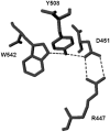
Figure 8. Hydrogen bonding between TPPI residues.
Interaction of residues W542, Y508, D451, and R447 through hydrogen bonding. X-ray crystallographic structure of TPPI, 3EDY, was modeled using the software DeepView v 4.01 with the hydrogen-bonding function activated. The hydrogen bridge formed by four amino acids, W542, Y508, D451, and R447, is shown by the dotted lines without the peptide backbone.
Figure 9
Figure 9. Interaction between Ca2+-binding amino acid residues and hydrogen-bonding quartet involving W542.
Interaction of residues W542, Y508, D451, and R447 through hydrogen bonding along with the Ca2+ binding residues.

Similar articles

Cited by

References

    1. Junaid MA, Pullarkat RK. Biochemistry of neuronal ceroid lipofuscinosis. Adv Genet. 2001;45:93–106. - PubMed
    1. Mole SE. The genetic spectrum of human neuronal ceroid-lipofuscinoses. Brain Pathol. 2004;14(1):70–76. - PMC - PubMed
    1. Golabek AA, Kida E. Tripeptidyl-peptidase I in health and disease. Biol Chem. 2006;387(8):1091–1099. - PubMed
    1. Zeman W, Donahue S, Dyken P, Green J. Leukodystrophies and poliodystrophies. In: Vinken PJ, Bruyn GW, editors. Handbook of Clinical Neurology. Amsterdam: Elsevier Science. 1970;Volume 10:588–679.
    1. Boustany R-M. Neurodystrophies and Neurolipidoses. In: Moser HW, editor. Handbook of Clinical Neurology. Amsterdam: Elsevier Science. 1996;Volume 22 (66):671–700.

Publication types

MeSH terms