. 2010 Aug 3;5(8):e11944.
doi: 10.1371/journal.pone.0011944.

IL-33 is produced by mast cells and regulates IgE-dependent inflammation

Affiliations

IL-33 is produced by mast cells and regulates IgE-dependent inflammation

Chia-Lin Hsu et al. PLoS One. .

Abstract

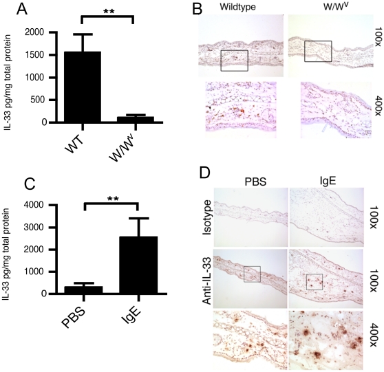
Background: IL-33 is a recently characterized IL-1 family cytokine and found to be expressed in inflammatory diseases, including severe asthma and inflammatory bowl disease. Recombinant IL-33 has been shown to enhance Th2-associated immune responses and potently increase mast cell proliferation and cytokine production. While IL-33 is constitutively expressed in endothelial and epithelial cells, where it may function as a transcriptional regulator, cellular sources of IL-33 and its role in inflammation remain unclear.

Methodology/principal findings: Here, we identify mast cells as IL-33 producing cells. IgE/antigen activation of bone marrow-derived mast cells or a murine mast cell line (MC/9) significantly enhanced IL-33. Conversely, recombinant IL-33 directly activated mast cells to produce several cytokines including IL-4, IL-5 and IL-6 but not IL-33. We show that expression of IL-33 in response to IgE-activation required calcium and that ionomycin was sufficient to induce IL-33. In vivo, peritoneal mast cells expressed IL-33 and IL-33 levels were significantly lower within the skin of mast cell deficient mice, compared to littermate controls. Local activation of mast cells promotes edema, followed by the recruitment of inflammatory cells. We demonstrate using passive cutaneous anaphylaxis, a mast cell-dependent model, that deficiency in ST2 or antibody blockage of ST2 or IL-33 ablated the late phase inflammatory response but that the immediate phase response was unaffected. IL-33 levels in the skin were significantly elevated only during the late phase.

Conclusions/significance: Our findings demonstrate that mast cells produce IL-33 after IgE-mediated activation and that the IL-33/ST2 pathway is critical for the progression of IgE-dependent inflammation.


Conflict of interest statement

Competing Interests: The authors have declared that no competing interests exist.

Figures

Figure 1
Figure 1. In vitro mast cells produce IL-33 upon specific activation by IgE.
1×10⁶ BMMC were primed with 1 µg/ml anti-DNP IgE (SPE-7 clone) overnight and stimulated by addition of 0.5 µg/ml DNP-HSA (black bars) or vehicle control (white bars). After the indicated times, gene expression of mouse IL-33 was determined by real-time RT-PCR (A). Either anti-DNP IgE or anti-OVA IgE was used in combination with either DNP-HSA or OVA, as indicated, and IL-33 gene expression determined (B). Intracellular staining for IL-33 was determined in permeabilized BMMC 24 hours after stimulation of 0.5 µg/ml DNP-HSA (C). Western blot analysis of BMMC lysate from stimulated or unstimulated cells 24 hours after stimulation (D). Recombinant, mouse IL-33109–266 was used as a positive control. Data represents 3 independent experiments. Primary mast cells were obtained by peritoneal lavage, identified as CD117+/FcεRI+ and intracellular staining for IL-33 performed after permeabilization (E). Data represents 2 independent experiments.
Figure 2
Figure 2. Mast cell expression of IL-33 requires calcium and is not induced by IL-33.
1×10⁶ BMMC were primed with 1 µg/ml anti-DNP IgE (SPE-7 clone) overnight and stimulated by addition of 0.5 µg/ml DNP-HSA or 10 ng/ml of recombinant IL-33109–266. After 4 hours, the gene expression of cytokines was determined by real-time RT-PCR in BMMC (A) and in MC/9 cells (B). IL-33 expression in response to ionomycin stimulation was determined using BMMC (C). The expression of IL-33 was then determined in BMMC after stimulation with 0.25 µM ionomycin or IgE/antigen in the presence or absence of 5 mM EDTA (D). * = p<0.05, ** = p<0.01 by Students t-test. Data represents the mean±SEM from 6 individual wells over two independent experiments.
Figure 3
Figure 3. ST2 is downregulated on mast cells after IgE-driven activation.
1×10⁶ BMMC (A) or MC/9 cells (B) were primed with 1 µg/ml anti-DNP IgE (SPE-7 clone) overnight and stimulated by addition of 0.5 µg/ml DNP-HSA or 10 ng/ml of recombinant IL-33109–266. After 4 hours, the gene expression of ST2 was determined by real-time RT-PCR. Surface expression of ST2 protein on BMMC was determined by flow cytometry after 6 hours (C). The levels of ST2 in supernatants were analyzed by ELISA after 6 hours (D). * = p<0.05, *** = p<0.005 by Students t-test. Data in A and C represents the mean±SEM from 3 to 6 individual wells over two independent experiments. Data in B is representative of results from three independent experiments.
Figure 4
Figure 4. In vivo mast cells constitutively express IL-33 and increase expression during anaphylaxis.
The tissue levels of IL-33 in the skin of wild type or mast cell deficient mice (W/Wv) were determined by ELISA (A). Additionally, frozen sections (7 µm) were stained for IL-33 immunoreactivity using biotinylated anti-IL-33 and ABC-DAB substrate and sections counterstained with hematoxylin (B). Total tissue IL-33 was determined in IgE-primed or sham skin in passive cutaneous anaphylaxis (C) and IL-33 immunoreactivity determined, as before (D). ** = p<0.01 by Students t-test. Data in panels A and C represents the mean±SEM from 5 individual mice while panels B and D are representative of results from 4 individual mice.
Figure 5
Figure 5. IL-33/ST2 pathway is critical for the development of IgE-driven tissue inflammation during anaphylaxis.
Induction of ear swelling during passive cutaneous anaphylaxis was determined after systemic antigen challenge in mice receiving no treatment, 10 µg anti-murine IL-33 antibody or 10 µg isotype control (A), anti-murine ST2 antibody or isotype control (B) or in ST2−/− or ST2−/+ control mice (C). Data represents the mean±SEM from 10–15 mice. The IL-33 protein levels in ear tissue from IgE injected or PBS injected sites were determined by ELISA during the PCA time course (D) (n = 3 mice per time point). Increases in IL-33 at IgE injected skin sites (determined as the delta between the PBS injected skin from the same individual) were determined in W/Wv, reconstituted W/Wv, ST2−/− or relevant control strains (n = 3–9 mice per group). * = p<0.05, ** = p<0.01, *** = p<0.005 by Students t-test.

Similar articles

Cited by

References

    1. Gilfillan AM, Rivera J. The tyrosine kinase network regulating mast cell activation. Immunol Rev. 2009;228:149–169. - PMC - PubMed
    1. Galli SJ, Nakae S, Tsai M. Mast cells in the development of adaptive immune responses. Nat Immunol. 2005;6:135–142. - PubMed
    1. Gilfillan AM, Tkaczyk C. Integrated signalling pathways for mast-cell activation. Nat Rev Immunol. 2006;6:218–230. - PubMed
    1. Schmitz J, Owyang A, Oldham E, Song Y, Murphy E, et al. IL-33, an interleukin-1-like cytokine that signals via the IL-1 receptor-related protein ST2 and induces T helper type 2-associated cytokines. Immunity. 2005;23:479–490. - PubMed
    1. Carriere V, Roussel L, Ortega N, Lacorre DA, Americh L, et al. IL-33, the IL-1-like cytokine ligand for ST2 receptor, is a chromatin-associated nuclear factor in vivo. Proc Natl Acad Sci U S A. 2007;104:282–287. - PMC - PubMed

Publication types

MeSH terms