. 2010 Jul 30;5(7):e11915.
doi: 10.1371/journal.pone.0011915.

Turning the table: plants consume microbes as a source of nutrients

Affiliations

Turning the table: plants consume microbes as a source of nutrients

Chanyarat Paungfoo-Lonhienne et al. PLoS One. .

Abstract

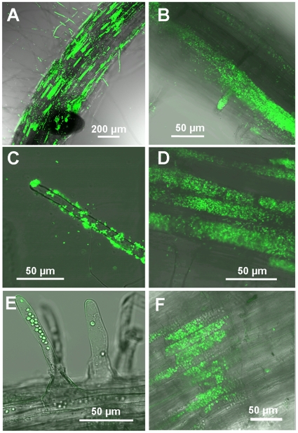
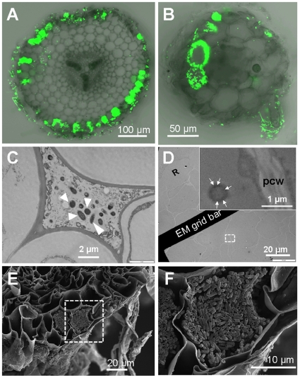
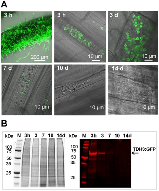
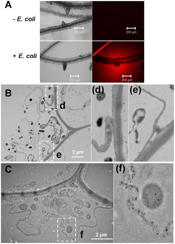
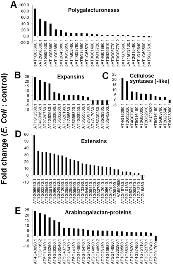
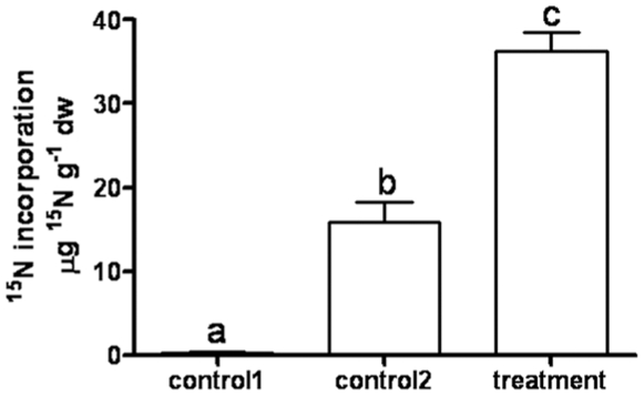
Interactions between plants and microbes in soil, the final frontier of ecology, determine the availability of nutrients to plants and thereby primary production of terrestrial ecosystems. Nutrient cycling in soils is considered a battle between autotrophs and heterotrophs in which the latter usually outcompete the former, although recent studies have questioned the unconditional reign of microbes on nutrient cycles and the plants' dependence on microbes for breakdown of organic matter. Here we present evidence indicative of a more active role of plants in nutrient cycling than currently considered. Using fluorescent-labeled non-pathogenic and non-symbiotic strains of a bacterium and a fungus (Escherichia coli and Saccharomyces cerevisiae, respectively), we demonstrate that microbes enter root cells and are subsequently digested to release nitrogen that is used in shoots. Extensive modifications of root cell walls, as substantiated by cell wall outgrowth and induction of genes encoding cell wall synthesizing, loosening and degrading enzymes, may facilitate the uptake of microbes into root cells. Our study provides further evidence that the autotrophy of plants has a heterotrophic constituent which could explain the presence of root-inhabiting microbes of unknown ecological function. Our discovery has implications for soil ecology and applications including future sustainable agriculture with efficient nutrient cycles.


Conflict of interest statement

Competing Interests: The authors have declared that no competing interests exist.

Figures

Figure 1
Figure 1. Roots of axenically grown Arabidopsis and tomato were incubated with E coli or yeast expressing green fluorescent protein (GFP E. coli or GFPyeast).
GFP E. coli was detected at the surface of roots and root hairs (A and C), and inside roots and root hairs (B and D). GFPYeast was present inside roots and root hairs (E and F). (A, D and F) and (B, C and E) correspond to tomato and Arabidopsis root, respectively. Fluorescent images were taken by confocal laser scanning microscopy (CLSM).
Figure 2
Figure 2. Root transverse sections and electron micrographs of tomato and Arabidopsis show GFP E. coli in the apoplast and inside root cells.
E. coli was detected inside tomato roots (A, C and D, E and F) and Arabidopsis roots (B). (A and B) Fluorescent images of transverse sectioned roots taken by CLSM. (C and D) Images taken by a transmission electron microscope. White triangles in (C) indicate E. coli cell present in apoplast. (D) Roots were probed with immunogold-labeled anti-GFP revealing E. coli in root cortex cells. Sub-image in (D) is a detail of dash-white square box. Gold labeling is marked with white arrows. Rhizodermis cell (R) and plant cell wall (pcw) is indicated. (F) is a detail image of (E) showing plant cells containing E. coli, and both images were taken by SEM.
Figure 3
Figure 3. Time course experiment of yeast degradation in tomato roots.
(A) The number of living yeast cells (fluorescing green) in tomato decreased over time as observed by CLSM. (B) The amount of recombinant TDH3:GFP protein present inside the roots 0decreased over time. Equal amounts of proteins from root extracts were separated by SDS-PAGE (B, Left) and analyzed by western blot using anti-GFP antibody to detect yeast recombinant TDH3:GFP protein (B, Right).
Figure 4
Figure 4. Root produced cellulase and extended the cell wall when incubated with E. coli Bl21.
(A) Incubation of Arabidopsis roots in 31 µg/mL resorufin cellubioside after incubating overnight with E. coli. After 2 h incubation, roots were viewed by CLSM. (B) TEM image of cell wall-like structure of plant roots encompassing bacteria. (C) TEM image of cellulase-gold labeling on the root sections with double labeling with the anti-GFP antibody. The size of the gold particle on bacteria is 15 nm (Au-particle specific to GFP E. coli) and gold particles on the plant material are 10 nm (Au-particle specific to plant cellulose). (d), (e) and (f) are detail images of insets d, e and f.
Figure 5
Figure 5. Arabidopsis genes involved in cell wall modification with differential expression at the time incubated with E coli Bl21 compared with control.
Gene expression more than 3 fold changes were shown. (A) Glycosyl hydrolases and lyases (x, c, p are the symbol for xyloglucan endotransglycosylase, cellulase and pectinases/putative pectinases, respectively). (B) Expansins. (C) Cellulose syntases and cellulose syntase-like. (D) Extensins. (E) Arabinogalactan proteins.
Figure 6
Figure 6. Incorporation of E. coli-derived 15N by leaves of tomato plants.
Roots of tomato grown in hydroponic culture were incubated with 15N-E. coli for 1 h. After washing of the roots, plants were further grown for 2 weeks. Then 2–3 new leaves were analyzed for 15N content. Control 1 are plants grown without 15N-E. coli. Control 2 are plants incubated for 2 h with filtered 15N-E. coli incubation solution. Results are depicted as mean ± SD (n = 7). Different letters indicate significant differences at p<0.001 (1-way ANOVA, Tukey's posthoc test).

References

    1. Baron C, Zambryski PC. Notes from the underground: Highlights from plant-microbe interactions. Trends Biotechnol. 1995;13:356–362.
    1. Boller T, He SY. Innate immunity in plants: an arms race between pattern recognition receptors in plants and effextors in microbial pathogens. Science. 2009;324:742–744. - PMC - PubMed
    1. Chisholm ST, Coaker G, Day B, Staskawicz BJ. Host-microbe interactions: Shaping the evolution of the plant immune response. Cell. 2006;124:803–814. - PubMed
    1. Reinhold-Hurek B, Hurek T. Life in grasses: diazotrophic endophytes. Trends Microbiol. 1998;6:139–144. - PubMed
    1. Triplett EW. Diazotrophic endophytes: Progress and prospects for nitrogen fixation in monocots. Plant Soil. 1996;186:29–38.

Publication types

MeSH terms