. 2010 Jul 30;5(7):e11917.
doi: 10.1371/journal.pone.0011917.

Retinoic acids potentiate BMP9-induced osteogenic differentiation of mesenchymal progenitor cells

Affiliations

Retinoic acids potentiate BMP9-induced osteogenic differentiation of mesenchymal progenitor cells

Wenli Zhang et al. PLoS One. .

Abstract

Background: As one of the least studied bone morphogenetic proteins (BMPs), BMP9 is one of the most osteogenic BMPs. Retinoic acid (RA) signaling is known to play an important role in development, differentiation and bone metabolism. In this study, we investigate the effect of RA signaling on BMP9-induced osteogenic differentiation of mesenchymal progenitor cells (MPCs).

Methodology/principal findings: Both primary MPCs and MPC line are used for BMP9 and RA stimulation. Recombinant adenoviruses are used to deliver BMP9, RARalpha and RXRalpha into MPCs. The in vitro osteogenic differentiation is monitored by determining the early and late osteogenic markers and matrix mineralization. Mouse perinatal limb explants and in vivo MPC implantation experiments are carried out to assess bone formation. We find that both 9CRA and ATRA effectively induce early osteogenic marker, such as alkaline phosphatase (ALP), and late osteogenic markers, such as osteopontin (OPN) and osteocalcin (OC). BMP9-induced osteogenic differentiation and mineralization is synergistically enhanced by 9CRA and ATRA in vitro. 9CRA and ATRA are shown to induce BMP9 expression and activate BMPR Smad-mediated transcription activity. Using mouse perinatal limb explants, we find that BMP9 and RAs act together to promote the expansion of hypertrophic chondrocyte zone at growth plate. Progenitor cell implantation studies reveal that co-expression of BMP9 and RXRalpha or RARalpha significantly increases trabecular bone and osteoid matrix formation.

Conclusion/significance: Our results strongly suggest that retinoid signaling may synergize with BMP9 activity in promoting osteogenic differentiation of MPCs. This knowledge should expand our understanding about how BMP9 cross-talks with other signaling pathways. Furthermore, a combination of BMP9 and retinoic acid (or its agonists) may be explored as effective bone regeneration therapeutics to treat large segmental bony defects, non-union fracture, and/or osteoporotic fracture.


Conflict of interest statement

Competing Interests: The authors have declared that no competing interests exist.

Figures

Figure 1
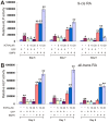
Figure 1. Retinoic acids induce osteogenic differentiation of mesenchymal progenitor cells (MPCs).
(A) 9-Cis-retinoic acid (9CRA) induces ALP activity in MPCs. Subconfluent C3H10T1/2 cells were treated with varying concentrations of 9CRA or solvent control. ALP activity was measured at the indicated time points. Each assay condition was carried out in triplicate in at least two independent batches of experiments. “*”, p<0.05; “**”, p<0.001 (vs. control groups). (B) All-trans-retinoic acid (ATRA) induces ALP activity in MPCs. C3H10T1/2 cells were treated with varying concentrations of ATRA or solvent control. ALP activity was measured at the indicated time points. Each assay condition was carried out in triplicate in at least two independent batches of experiments. “*”, p<0.01; “**”, p<0.001 (vs. control groups).
Figure 2
Figure 2. Retinoic acids and BMP9 act synergistically in inducing ALP activity in MPCs.
(A) 9CRA induces ALP activity in MPCs. Subconfluent MEFs were infected with AdBMP9 (MOI = 5) or AdGFP (MOI = 5), followed by treatment with varying concentrations of 9CRA or solvent control. ALP activity was measured at the indicated time points. Each assay condition was carried out in triplicate in at least two independent batches of experiments. “*”, p<0.05; “**”, p<0.001 (vs. control groups). (B) ATRA induces ALP activity in MPCs. MEFs were infected with AdBMP9 (MOI = 5) or AdGFP (MOI = 5), followed by treatment with varying concentrations of ATRA or solvent control. ALP activity was measured at the indicated time points. Each assay condition was carried out in triplicate in at least two independent batches of experiments. “*”, p<0.05; “**”, p<0.001 (vs. control groups).
Figure 3
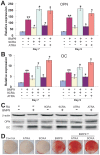
Figure 3. Retinoids potentiate BMP9-induced late osteogenic markers and matrix mineralization in MPCs.
(A) qPCR analysis of retinoic acids and BMP9 induced osteopontin (OPN) expression. Subconfluent MEFs were infected with AdBMP9 or AdGFP (i.e., -BMP9 groups, MOI = 5), and then treated with 9CRA (20 µM), ATRA (20 µM), or solvent control. At day 7 and day 9, the cells were collected for total RNA isolation. RNA was subjected to RT-PCR transcription, which was used as templates for qPCR analysis using primers specific for mouse OPN. Each assay condition was carried out in triplicate. All samples were normalized using endogenous levels of GAPDH. “*”, p<0.05; “**”, p<0.01 (vs. control groups). (B) qPCR analysis of retinoic acids and BMP9 induced osteocalcin (OC) expression. Samples prepared in (A) were used for qPCR analysis using primers specific for mouse OC. Each assay condition was carried out in triplicate. “*”, p<0.05; “**”, p<0.01 (vs. control groups). (C) Western blotting analysis of retinoic acids and BMP9 induced OPN and OC expression. MEFs were infected with AdBMP9 or AdGFP (i.e., -BMP9 groups, MOI = 5), and then treated with 9CRA (20 µM), ATRA (20 µM), or solvent control. At day 7, cells were lysed and subjected to Western blotting analysis using anti-OPN or anti-OC antibody (Santa Cruz Biotechnology). Anti-β actin antibody was used to demonstrate equal loading of all samples. (D) Retinoic acids and BMP9 induce matrix mineralization. MEFs were infected with AdBMP9 or AdGFP (i.e., -BMP9 groups, MOI = 5), and then treated with 9CRA (20 µM), ATRA (20 µM), or solvent control. At day 14, cells were subjected to Alizarin Red S staining. Experiments were carried out in duplicate and representative results are shown.
Figure 4
Figure 4. Retinoids induce BMP9 expression and activate BMP-Smad pathway.
(A) Retinods induce BMP9 expression in MPCs. Subconfluent MEFs were treated with 9CRA (20 µM), ATRA (20 µM), or solvent control. At day 3 and day 5, the cells were collected for total RNA isolation. RNA was subjected to RT-PCR transcription, which was used as templates for qPCR analysis using primers specific for mouse BMP9. Each assay condition was carried out in triplicate. All samples were normalized using endogenous levels of GAPDH. “*”, p<0.05; “**”, p<0.01 (vs. control groups). (B) Retinods activate BMPR Smad reporter and Runx2 reporter activity in MPCs. MEFs were transfected with BMPR Smad reporter, p12xSBE-Luc or OC promoter containing Runx2-responsive element reporter, p6xOSE-Luc. The transfected cells were replated at 16h after transfection, followed by a treatment with 9CRA (20 µM), ATRA (20 µM), or solvent control. 48 h after treatment, the cells were lyzed for luciferase activity assay using Promega's Luciferase Assay kit. Each assay condition was carried out in triplicate. Luciferase activity was normalized by total cellular protein concentrations among the samples. “*”, p<0.05; “**”, p<0.001 (vs. control groups).
Figure 5
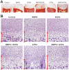
Figure 5. Retinoids and BMP9 promote the expansion of hypertrophic chondrocyte zone in organ culture.
(A) Harvest, transduction and labeling of mouse E18.5 forelimbs (n = 5 each group). E18.5 forelimbs were dissected, and the skin was removed with the soft tissues attached. Recombinant adenovirus (5×10∧10 pfu in 1 ml medium) expressing AdGFP or AdBMP9 added to culture medium, with or without 9CRA (20 µM) or ATRA (20 µM). After two weeks, the cultured limbs were harvested, embedded, and subjected to Alizarin Red S stating. Representative low magnification images are shown. (B) Histologic evaluation. The above samples were subjected to sectioning and H & E staining, and recorded under bright field with 400× magnification. The approximate lengths of hypertrophic zones were indicated. Representative images are shown. HC, hypertrophic chondrocyte zone.
Figure 6
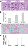
Figure 6. Retinoids enhance BMP9-induced ectopic ossification.
(A) Histological evaluation of ectopic bone formation. Subconfluent MEFs were co-infected with AdBMP9 and AdRFP, AdR-RXRα, or AdR-RARα adenoviruses (MOI = 10) for 16 h. The infected cells were implanted subcutaneously (5×10⁶/injection) into the flanks of athymic nude (nu/nu) mice (5 mice/group, 4–6 week old, male, Harlan Sprague Dawley). At 4 wk after implantation, animals were sacrificed, and the implantation sites were retrieved, fixed and decalcified. The paraffin-embedded sections were subjected to H & E staining. Representative images are shown. BM, bone matrix (ossified and osteoid); Ob, osteoblast. (B) Quantitative analysis of trabecular and osteoid matrix area. The average trabecular bone and osteoid matrix areas were determined. At least 10 samples (with ×100 magnification) from each group were randomly selected and analyzed by using ImageJ software. “*” p<0.05, “**” p<0.001. (C) Masson's Trichrome staining of ectopic bone masses. Tissue sections prepared in were subjected to Masson's Trichrome staining. Representative images are shown. Magnification, ×400. (D) Quantitative analysis of % trabecular/osteoid area over total area was done by using ImageJ. At least 10 samples (with ×100 magnification) from each group were randomly selected and analyzed. “*” p<0.01.

Similar articles

Cited by

References

    1. Sacchetti B, Funari A, Michienzi S, Di Cesare S, Piersanti S, et al. Self-renewing osteoprogenitors in bone marrow sinusoids can organize a hematopoietic microenvironment. Cell. 2007;131:324–336. - PubMed
    1. Prockop DJ. Marrow stromal cells as stem cells for nonhematopoietic tissues. Science. 1997;276:71–74. - PubMed
    1. Pittenger MF, Mackay AM, Beck SC, Jaiswal RK, Douglas R, et al. Multilineage potential of adult human mesenchymal stem cells. Science. 1999;284:143–147. - PubMed
    1. Aubin JE. Advances in the osteoblast lineage. Biochem Cell Biol. 1998;76:899–910. - PubMed
    1. Deng ZL, Sharff KA, Tang N, Song WX, Luo J, et al. Regulation of osteogenic differentiation during skeletal development. Front Biosci. 2008;13:2001–2021. - PubMed

Publication types

MeSH terms