

Glypican-1, phosphacan/receptor protein-tyrosine phosphatase-ζ/β and its ligand, tenascin-C, are expressed by neural stem cells and neural cells derived from embryonic stem cells
- PMID: 20689858
- PMCID: PMC2914431
- DOI: 10.1042/AN20100001
Glypican-1, phosphacan/receptor protein-tyrosine phosphatase-ζ/β and its ligand, tenascin-C, are expressed by neural stem cells and neural cells derived from embryonic stem cells
Abstract
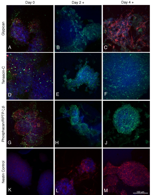
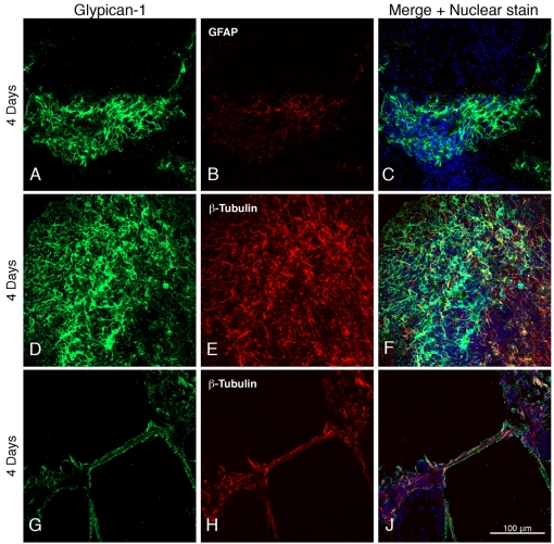
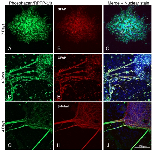
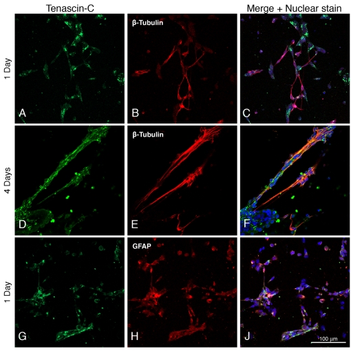
The heparan sulfate proteoglycan glypican-1, the chondroitin sulfate proteoglycan phosphacan/RPTP (receptor protein-tyrosine phosphatase)-zeta/beta and the extracellular matrix protein tenascin-C were all found to be expressed by neural stem cells and by neural cells derived from them. Expression of proteoglycans and tenascin-C increased after retinoic acid induction of SSEA1-positive ES (embryonic stem) cells to nestin-positive neural stem cells, and after neural differentiation, proteoglycans and tenascin-C are expressed by both neurons and astrocytes, where they surround cell bodies and processes and in certain cases show distinctive expression patterns. With the exception of tenascin-C (whose expression may decrease somewhat), expression levels do not change noticeably during the following 2 weeks in culture. The significant expression, by neural stem cells and neurons and astrocytes derived from them, of two major heparan sulfate and chondroitin sulfate proteoglycans of nervous tissue and of tenascin-C, a high-affinity ligand of phosphacan/RPTP-zeta/beta, indicates that an understanding of their specific functional roles in stem cell neurobiology will be important for the therapeutic application of this new technology in facilitating nervous tissue repair and regeneration.
Keywords: astrocyte; glypican; heparan sulfate; neuron; phosphacan; proteoglycan.
Figures
References
-
- Auerbach W, Dunmore JH, Fairchild-Huntress V, Fang Q, Auerbach AB, Huszar D, Joyner AL. Establishment and chimera analysis of 129/SvEv- and C57BL/6-derived mouse embryonic stem cell lines. BioTechniques. 2000;29:1024–1032. - PubMed
-
- Bain G, Yao M, Huettner J, Finley M, Gottlieb D. Neuronlike cells derived in culture from P19 embryonal carcinoma and embryonic stem cells. In: Banker G, Goslin K, editors. In Culturing Nerve Cells. MIT, Cambridge, MA: 1998. pp. 189–212.
-
- Beckett K, Franch-Marro X, Vincent JP. Glypican-mediated endocytosis of Hedgehog has opposite effects in flies and mice. Trends Cell Biol. 2008;18:360–363. - PubMed
-
- Bourdon MA, Coleman RE, Blasberg RG, Groothius DR, Bigner DD. Monoclonal antibody localization in subcutaneous and intracranial human glioma zenografts: paired-label and imaging analysis. Anticancer Res. 1985;4:133–140. - PubMed
-
- Braam SR, Denning C, van den Brink S, Kats P, Hochstenbach R, Passier R, Mummery CL. Improved genetic manipulation of human embryonic stem cells. Nat Methods. 2008;5:389–392. - PubMed
Publication types
MeSH terms
Substances
Grants and funding
LinkOut - more resources
Full Text Sources