Female germ unit in Genlisea and Utricularia, with remarks about the evolution of the extra-ovular female gametophyte in members of Lentibulariaceae
- PMID: 20689973
- PMCID: PMC3066386
- DOI: 10.1007/s00709-010-0185-x
Female germ unit in Genlisea and Utricularia, with remarks about the evolution of the extra-ovular female gametophyte in members of Lentibulariaceae
Abstract
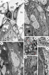
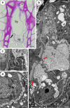
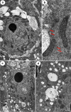
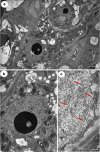
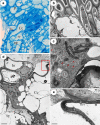
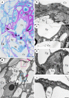
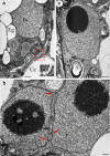
Lentibulariaceae is the largest family among carnivorous plants which displays not only an unusual morphology and anatomy but also the special evolution of its embryological characteristics. It has previously been reported by authors that Utricularia species lack a filiform apparatus in the synergids. The main purposes of this study were to determine whether a filiform apparatus occurs in the synergids of Utricularia and its sister genus Genlisea, and to compare the female germ unit in these genera. The present studies clearly show that synergids in both genera possess a filiform apparatus; however, it seems that Utricularia quelchii synergids have a simpler structure compared to Genlisea aurea and other typical angiosperms. The synergids are located at the terminal position in the embryo sacs of Pinguicula, Genlisea and were probably also located in that position in common Utricularia ancestor. This ancestral characteristic still occurs in some species from the Bivalvaria subgenus. An embryo sac, which grows out beyond the limit of the integument and has contact with nutritive tissue, appeared independently in different Utricularia lineages and as a consequence of this, the egg apparatus changes position from apical to lateral.
Figures
Similar articles
-
Actin cytoskeleton in the extra-ovular embryo sac of Utricularia nelumbifolia (Lentibulariaceae).Protoplasma. 2012 Jul;249(3):663-70. doi: 10.1007/s00709-011-0306-1. Epub 2011 Jul 24. Protoplasma. 2012. PMID: 21786167 Free PMC article.
-
Functional anatomy of the ovule in Genlisea with remarks on ovule evolution in Lentibulariaceae.Protoplasma. 2009 Jul;236(1-4):39-48. doi: 10.1007/s00709-009-0045-8. Epub 2009 May 13. Protoplasma. 2009. PMID: 19437102
-
The Chloroplast Genome of Utricularia reniformis Sheds Light on the Evolution of the ndh Gene Complex of Terrestrial Carnivorous Plants from the Lentibulariaceae Family.PLoS One. 2016 Oct 20;11(10):e0165176. doi: 10.1371/journal.pone.0165176. eCollection 2016. PLoS One. 2016. PMID: 27764252 Free PMC article.
-
Recent progress in understanding the evolution of carnivorous lentibulariaceae (lamiales).Plant Biol (Stuttg). 2006 Nov;8(6):748-57. doi: 10.1055/s-2006-924706. Plant Biol (Stuttg). 2006. PMID: 17203430 Review.
-
Evolution of unusual morphologies in Lentibulariaceae (bladderworts and allies) and Podostemaceae (river-weeds): a pictorial report at the interface of developmental biology and morphological diversification.Ann Bot. 2016 Apr;117(5):811-32. doi: 10.1093/aob/mcv172. Epub 2015 Nov 20. Ann Bot. 2016. PMID: 26589968 Free PMC article. Review.
Cited by
-
Unusual developmental morphology and anatomy of vegetative organs in Utricularia dichotoma-leaf, shoot and root dynamics.Protoplasma. 2020 Mar;257(2):371-390. doi: 10.1007/s00709-019-01443-6. Epub 2019 Oct 28. Protoplasma. 2020. PMID: 31659470 Free PMC article.
-
Integument cell differentiation in dandelions (Taraxacum, Asteraceae, Lactuceae) with special attention paid to plasmodesmata.Protoplasma. 2016 Sep;253(5):1365-72. doi: 10.1007/s00709-015-0894-2. Epub 2015 Oct 10. Protoplasma. 2016. PMID: 26454638 Free PMC article.
-
Actin cytoskeleton in the extra-ovular embryo sac of Utricularia nelumbifolia (Lentibulariaceae).Protoplasma. 2012 Jul;249(3):663-70. doi: 10.1007/s00709-011-0306-1. Epub 2011 Jul 24. Protoplasma. 2012. PMID: 21786167 Free PMC article.
-
Syncytia in Utricularia: Origin and Structure.Results Probl Cell Differ. 2024;71:143-155. doi: 10.1007/978-3-031-37936-9_8. Results Probl Cell Differ. 2024. PMID: 37996677
-
Atypical DNA methylation, sRNA-size distribution, and female gametogenesis in Utricularia gibba.Sci Rep. 2021 Aug 3;11(1):15725. doi: 10.1038/s41598-021-95054-y. Sci Rep. 2021. PMID: 34344949 Free PMC article.
References
-
- Adamec L. Mineral nutrient relations in the aquatic carnivorous plant Utricularia australis and its investment in carnivory. Fund Appl Limnol. 2008;171:175–183. doi: 10.1127/1863-9135/2008/0171-0175. - DOI
-
- Begum M. Studies on the embryology of Utricularia graminifolia, Vahl. Curr Sci. 1965;34:355–356.
Publication types
MeSH terms
LinkOut - more resources
Full Text Sources

