

Review
doi: 10.1146/annurev-physiol-042210-121137.
Role of tissue factor in venous thrombosis
Affiliations
- PMID: 20690821
- PMCID: PMC3076951
- DOI: 10.1146/annurev-physiol-042210-121137
Item in Clipboard
Review
Role of tissue factor in venous thrombosis
Annu Rev Physiol.
2011.
Abstract
Venous thromboembolism (VTE) is a leading cause of morbidity and mortality worldwide. However, the mechanisms by which clots are formed in the deep veins have not been determined. Tissue factor (TF) is the primary initiator of the coagulation cascade and is essential for hemostasis. Under pathological conditions, TF is released into the circulation on small-membrane vesicles termed microparticles (MPs). Recent studies suggest that elevated levels of MP TF may trigger thrombosis. This review provides an overview of the role of TF in VTE.
Figures
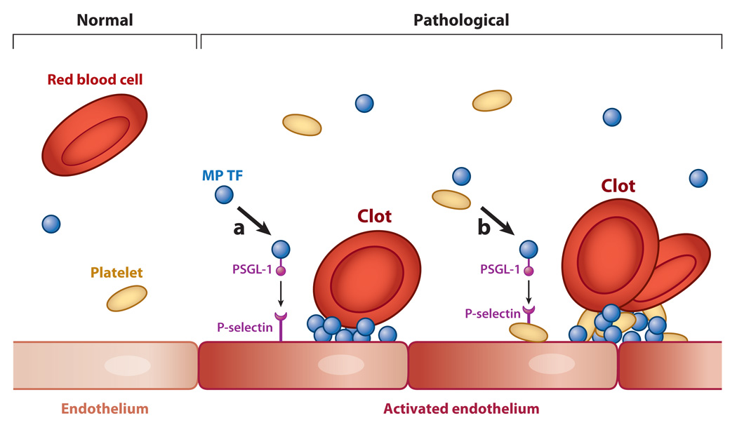
Formation of a venous clot. Venous thrombi are fibrin-rich clots that develop in the absence of gross endothelial damage. In a healthy vein (left), high levels of tissue factor pathway inhibitor (TFPI), thrombomodulin (TM), and endothelial cell protein C receptor (EPCR) maintain an antithrombotic phenotype (normal endothelium). In pathological conditions (right), elevated levels of tissue factor (TF)-positive microparticles (MPs) are present in the blood, and reduced blood flow, activation of the venous endothelium, and deposition of platelets may conspire to trigger formation of a thrombotic clot. Here we present two potential mechanisms that initiate activation of the coagulation system: (a) TF-positive MPs expressing P-selectin glycoprotein-1 (PSGL-1) dock to an activated endothelium expressing P-selectin, and (b) TF-positive MPs bind to activated platelets that adhere to the activated endothelium. In both cases, the presence of MP TF serves as a potent trigger of coagulation activation, and the continuous delivery of TF-positive MPs may enhance propagation of the thrombus.
Similar articles
-
Microparticles in hemostasis and thrombosis.Circ Res. 2011 May 13;108(10):1284-97. doi: 10.1161/CIRCRESAHA.110.233056. Circ Res. 2011. PMID: 21566224 Free PMC article. Review.
-
Tissue factor expressed by circulating cancer cell-derived microparticles drastically increases the incidence of deep vein thrombosis in mice.J Thromb Haemost. 2015 Jul;13(7):1310-9. doi: 10.1111/jth.13002. Epub 2015 Jun 8. J Thromb Haemost. 2015. PMID: 25955268 Free PMC article.
-
Tumor-derived tissue factor-positive microparticles and venous thrombosis in cancer patients.Blood. 2013 Sep 12;122(11):1873-80. doi: 10.1182/blood-2013-04-460139. Epub 2013 Jun 24. Blood. 2013. PMID: 23798713 Free PMC article. Review.
-
Human pancreatic tumors grown in mice release tissue factor-positive microvesicles that increase venous clot size.J Thromb Haemost. 2017 Nov;15(11):2208-2217. doi: 10.1111/jth.13809. Epub 2017 Sep 20. J Thromb Haemost. 2017. PMID: 28834179
-
Microparticle-associated tissue factor activity in patients with acute unprovoked deep vein thrombosis and during the course of one year.Thromb Res. 2014 Nov;134(5):1093-6. doi: 10.1016/j.thromres.2014.07.041. Epub 2014 Aug 22. Thromb Res. 2014. PMID: 25262106
Cited by
-
Recent advances in understanding, diagnosing and treating venous thrombosis.F1000Res. 2020 Oct 6;9:F1000 Faculty Rev-1206. doi: 10.12688/f1000research.27115.1. eCollection 2020. F1000Res. 2020. PMID: 33082930 Free PMC article. Review.
-
In silico analyses of blood flow and oxygen transport in human micro-veins and valves.Clin Hemorheol Microcirc. 2022;81(1):81-96. doi: 10.3233/CH-211345. Clin Hemorheol Microcirc. 2022. PMID: 35034895 Free PMC article.
-
Interplay between alternatively spliced Tissue Factor and full length Tissue Factor in modulating coagulant activity of endothelial cells.Thromb Res. 2017 Aug;156:1-7. doi: 10.1016/j.thromres.2017.05.028. Epub 2017 May 25. Thromb Res. 2017. PMID: 28570958 Free PMC article.
-
Mice with Reduced PAR4 Reactivity show Decreased Venous Thrombosis and Platelet Procoagulant Activity.bioRxiv [Preprint]. 2024 Oct 17:2024.10.14.617127. doi: 10.1101/2024.10.14.617127. bioRxiv. 2024. Update in: J Thromb Haemost. 2025 Apr;23(4):1278-1288. doi: 10.1016/j.jtha.2024.12.031. PMID: 39463946 Free PMC article. Updated. Preprint.
-
A low balance between microparticles expressing tissue factor pathway inhibitor and tissue factor is associated with thrombosis in Behçet's Syndrome.Sci Rep. 2016 Dec 7;6:38104. doi: 10.1038/srep38104. Sci Rep. 2016. PMID: 27924945 Free PMC article.
References
-
- Mackman N, Tilley RE, Key NS. Role of the extrinsic pathway of blood coagulation in hemostasis and thrombosis. Arterioscler. Thromb. Vasc. Biol. 2007;27(8):1687–1693. - PubMed
-
- Komiyama Y, Pedersen AH, Kisiel W. Proteolytic activation of human factors IX and X by recombinant human factor VIIa: effects of calcium, phospholipids, and tissue factor. Biochemistry. 1990;29(40):9418–9425. - PubMed
-
- Fleck RA, Rao LV, Rapaport SI, Varki N. Localization of human tissue factor antigen by immunostaining with monospecific, polyclonal antihuman tissue factor antibody. Thromb. Res. 1990;59(2):421–437. - PubMed
Publication types
MeSH terms
Substances
Grants and funding
LinkOut - more resources
Full Text Sources
Other Literature Sources
Medical
Miscellaneous
