

Kawasaki disease: novel insights into etiology and genetic susceptibility
- PMID: 20690826
- PMCID: PMC3021097
- DOI: 10.1146/annurev-med-042409-151944
Kawasaki disease: novel insights into etiology and genetic susceptibility
Abstract
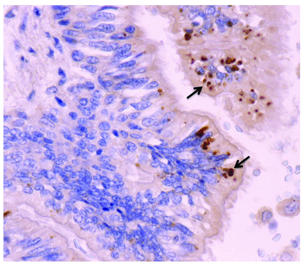
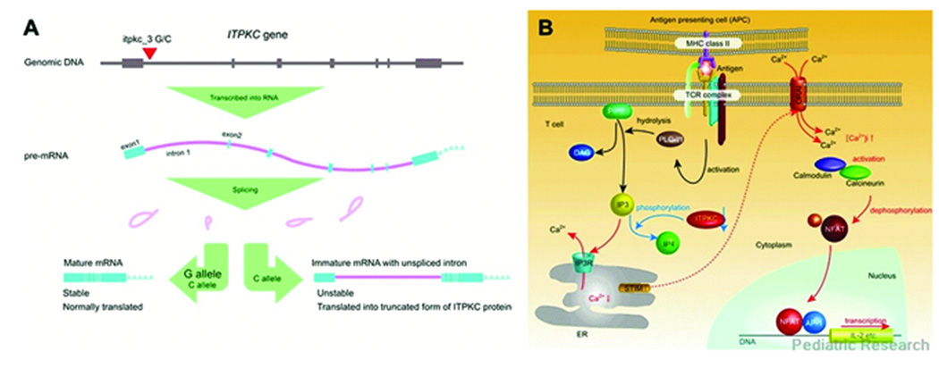
Kawasaki disease (KD) is a vasculitis of young childhood that particularly affects the coronary arteries. Molecular analysis of the oligoclonal IgA response in acute KD led to production of synthetic KD antibodies. These antibodies identify intracytoplasmic inclusion bodies in acute KD tissues. Light and electron microscopic studies indicate that the inclusion bodies are consistent with aggregates of viral proteins and RNA. Advances in molecular genetic analysis and completion of the Human Genome Project have sparked a worldwide effort to identify genes associated with KD. A polymorphism of one such gene, ITPKC, a negative regulator of T cell activation, confers susceptibility to KD in Japanese populations and increases the risk of developing coronary artery abnormalities in both Japanese and U.S. children. Identification of the etiologic agent and of genes conferring KD susceptibility are the best means of improving diagnosis and therapy and enabling prevention of this important disorder of childhood.
Figures
Similar articles
-
Genetic polymorphisms in Kawasaki disease.Acta Pharmacol Sin. 2011 Oct;32(10):1193-8. doi: 10.1038/aps.2011.93. Epub 2011 Sep 5. Acta Pharmacol Sin. 2011. PMID: 21892198 Free PMC article. Review.
-
ITPKC functional polymorphism associated with Kawasaki disease susceptibility and formation of coronary artery aneurysms.Nat Genet. 2008 Jan;40(1):35-42. doi: 10.1038/ng.2007.59. Epub 2007 Dec 16. Nat Genet. 2008. PMID: 18084290 Free PMC article.
-
Single-nucleotide polymorphism rs7251246 in ITPKC is associated with susceptibility and coronary artery lesions in Kawasaki disease.PLoS One. 2014 Mar 12;9(3):e91118. doi: 10.1371/journal.pone.0091118. eCollection 2014. PLoS One. 2014. PMID: 24621571 Free PMC article.
-
Immunogenetics of Kawasaki disease.Clin Rev Allergy Immunol. 2020 Aug;59(1):122-139. doi: 10.1007/s12016-020-08783-9. Clin Rev Allergy Immunol. 2020. PMID: 32200494 Review.
-
Susceptibility genes for Kawasaki disease: toward implementation of personalized medicine.J Hum Genet. 2009 Feb;54(2):67-73. doi: 10.1038/jhg.2008.9. Epub 2009 Jan 16. J Hum Genet. 2009. PMID: 19158812 Review.
Cited by
-
Systematic confirmation study of GWAS-identified genetic variants for Kawasaki disease in a Chinese population.Sci Rep. 2015 Feb 3;5:8194. doi: 10.1038/srep08194. Sci Rep. 2015. PMID: 25645453 Free PMC article.
-
Association between Kawasaki Disease and Prenatal Exposure to Ambient and Industrial Air Pollution: A Population-Based Cohort Study.Environ Health Perspect. 2020 Oct;128(10):107006. doi: 10.1289/EHP6920. Epub 2020 Oct 19. Environ Health Perspect. 2020. PMID: 33074736 Free PMC article.
-
Prediction of Immune Infiltration Diagnostic Gene Biomarkers in Kawasaki Disease.J Immunol Res. 2022 Jun 17;2022:8739498. doi: 10.1155/2022/8739498. eCollection 2022. J Immunol Res. 2022. PMID: 35755167 Free PMC article.
-
Kawasaki Disease: A Clinician's Update.Int J Pediatr. 2013;2013:645391. doi: 10.1155/2013/645391. Epub 2013 Oct 27. Int J Pediatr. 2013. PMID: 24282419 Free PMC article. Review.
-
Tropospheric winds from northeastern China carry the etiologic agent of Kawasaki disease from its source to Japan.Proc Natl Acad Sci U S A. 2014 Jun 3;111(22):7952-7. doi: 10.1073/pnas.1400380111. Epub 2014 May 19. Proc Natl Acad Sci U S A. 2014. PMID: 24843117 Free PMC article.
References
-
- Newburger JW, Takahashi M, Burns JC, et al. The treatment of Kawasaki syndrome with intravenous gamma globulin. N Engl J Med. 1986;315:341–347. - PubMed
-
- Newburger JW, Sleeper LA, McCrindle BW, et al. Randomized trial of pulsed corticosteroid therapy for primary treatment of Kawasaki disease. N Engl J Med. 2007;356:663–675. - PubMed
-
- Kawasaki T. Acute febrile mucocutaneous syndrome with lymphoid involvement with specific desquamation of the fingers and toes in children. Arerugi. 1967;16:178–222. - PubMed
-
- Patriarca PA, Rogers MF, Morens DM, et al. Kawasaki syndrome: association with the application of rug shampoo. Lancet. 1982;2:578–580. - PubMed
-
- Ohga K, Yamanaka R, Kinumaki H, et al. Kawasaki disease and rug shampoo. Lancet. 1983;1:930. - PubMed
Publication types
MeSH terms
Substances
Grants and funding
LinkOut - more resources
Full Text Sources
Medical
Miscellaneous
