. 2010 Aug 6:10:84.
doi: 10.1186/1471-213X-10-84.

Involvement of the Reck tumor suppressor protein in maternal and embryonic vascular remodeling in mice

Affiliations

Involvement of the Reck tumor suppressor protein in maternal and embryonic vascular remodeling in mice

Ediriweera P S Chandana et al. BMC Dev Biol. .

Abstract

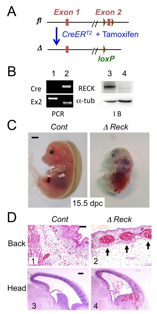
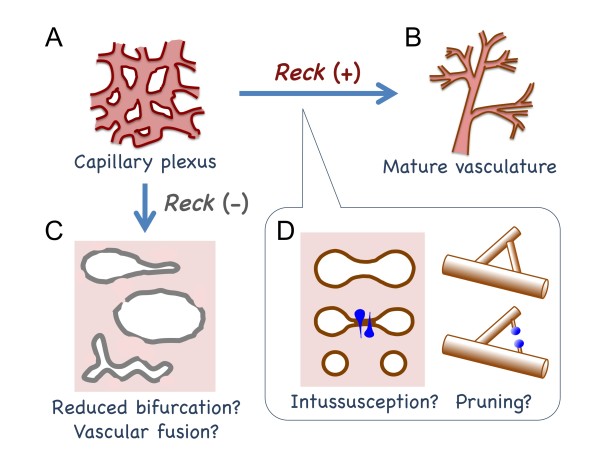
Background: Developmental angiogenesis proceeds through multiple morphogenetic events including sprouting, intussusception, and pruning. Mice lacking the membrane-anchored metalloproteinase regulator Reck die in utero around embryonic day 10.5 with halted vascular development; however, the mechanisms by which this phenotype arises remain unclear.

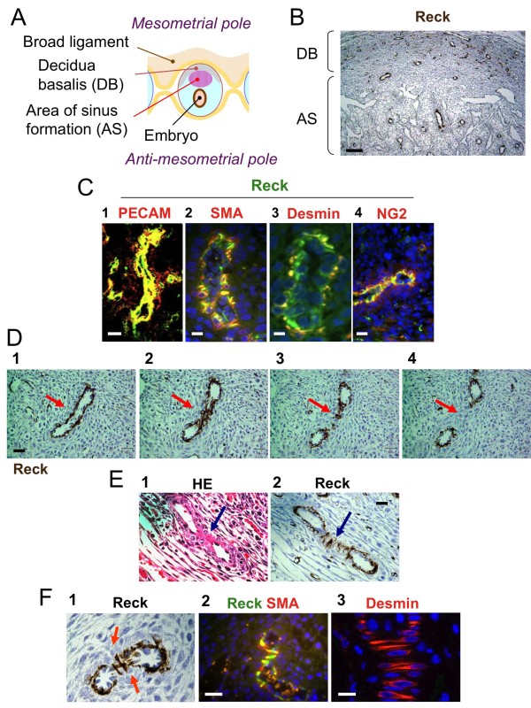
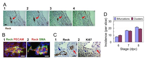
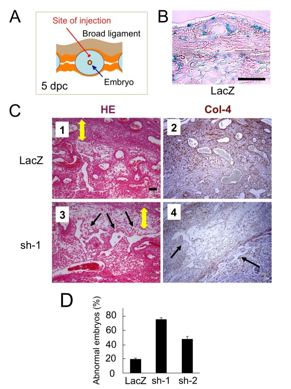
Results: We found that Reck is abundantly expressed in the cells associated with blood vessels undergoing angiogenesis or remodelling in the uteri of pregnant female mice. Some of the Reck-positive vessels show morphological features consistent with non-sprouting angiogenesis. Treatment with a vector expressing a small hairpin RNA against Reck severely disrupts the formation of blood vessels with a compact, round lumen. Similar defects were found in the vasculature of Reck-deficient or Reck conditional knockout embryos.

Conclusions: Our findings implicate Reck in vascular remodeling, possibly through non-sprouting angiogenesis, in both maternal and embyonic tissues.


Figures

Figure 1
Figure 1
Reck-immunoreactivity associated with blood vessels in the mouse implantation chamber. (A) Distinct domains in the mouse implantation chamber at around 7 dpc. (B) Reck-immunoreactivity (dark brown) in the AS and DB in a longitudinal section of a 7-dpc mouse implantation chamber. (C) Loop-shaped structures in DB sections doubly stained for Reck (green) and an endothelial cell marker (red) [PECAM; panel 1] or a mural cell marker (red) [SMA, desmin, or NG2; panels 2, 3, 4, respectively] followed by nuclear counter-staining with DAPI (blue signals; panels 2-4). (D) An example of bifurcating vessels in the DB found in serial sections (4 μm-thick) stained for Reck. Red arrows indicate protruding vessel walls (panel 1) which form a contact zone (panel 2) and eventually separate the vessel into two smaller tubes (panels 3, 4). (E) An example of characteristic Reck-positive cells associated with the contact zone. Two adjacent sections were stained with hematoxylin-eosin (H&E) (panel 1) and immuno-stained for Reck (panel 2), respectively. Blue arrows indicate the contact zone. (F) The wedge-shaped cells lying across the contact zone are positive for Reck (panel 1), SMA (panel 2; fluorescent double staining with Reck), and desmin (panel 3; fluorescent staining). Scale bar: B, 100 μm; C, E, F, 20 μm; D, 30 μm.
Figure 2
Figure 2
Reck-immunoreactivity associated with clusters of vascular cells in the mouse implantation chamber. (A) An example of a cluster of cells (red arrow) positive for Reck at a vessel terminus found in the DB. (B) Immunofluorescent double staining of a terminal cell cluster for Reck (green) and PECAM (red; panel 1) or Reck (red) and SMA (green; panel 2) followed by nuclear counter-staining with DAPI (blue). (C) Two adjacent sections stained for Reck (panel 1) and Ki67 (panel 2), respectively. Blue arrows: a bifurcating vessel. Red arrows: a terminal cell cluster. (D) Temporal changes in the number of Reck-positive bifurcating vessels (blue bar) and terminal cell clusters (red bar) per slice from the medial part of implantation chambers. Bar represents mean ± s.e.m., n = 7. The difference between stages is statistically significant (p < 0.05) for both Reck-positive bifurcating vessels and terminal cell clusters. Scale bar: A, 30 μm; B, C, 20 μm.
Figure 3
Figure 3
Reck shRNA interferes with vascular remodeling in the mouse implantation chamber. (A) The site of bead injection (red line) in the mesometrial area at 5 dpc. (B) Control experiments to assess the efficiency of gelatin-bead-mediated gene transfer into the implantation chamber. Cationized gelatin beads impregnated with a LacZ-expression vector were injected into the mesometrial area at 5 dpc, and tissue slices were prepared at 10 dpc and stained with X-gal. A typical image is shown. (C) Mesometrial tissue at 10 dpc that had been transfected on 5 dpc with a plasmid expressing either LacZ (panels 1, 2) or shRNA against Reck (sh-1; panels 3, 4) was sliced and stained with H&E (panels 1, 3) or immunostained for type IV collagen (panels 2, 4). The yellow double-headed arrows indicate the DB, and the black arrows abnormal vessels. (D) The frequency of samples with abnormal decidua after transfection with vectors expressing LacZ or either of the two shRNA against Reck, sh-1 and sh-2, with different efficacy. Embryos were scored abnormal when the vessels in the transfected areas were severely disrupted as shown in panels 3 and 4 in C; the abnormality was often accompanied by reduced cellularity in the areas as well. Bar represents mean ± s.e.m., n = 7 pregnant mice. Total implantation chambers tested: LacZ, 81; sh-1, 88; sh-2, 86. Student's t-test: LacZ vs. sh-1, p = 1.7 × 10⁻⁸; LacZ vs. sh-2, p = 1.4 × 10⁻⁵; sh-1 vs. sh-2, p = 1.6 × 10⁻⁵. Scale bar: B, 200 μm; C, 100 μm.
Figure 4
Figure 4
Vascular defects in Reck-deficient mice. (A) Schematic representation of the 5'-terminal region of the wild type (wt) and a Reck mutant (Reck-) allele. This mutant allele lacks exon 1 and hence no Reck protein is expressed [15]. (B) A typical E10.5 wild type embryo (panel 1) and a Reck-/- embryo (panel 2) which shows abdominal hemorrhage and dilated vessels (panel 3, magnified view of the area indicated by yellow box in panel 2). The heart was beating in this typical mutant embryo. (C) The perineural area in the sagittal sections of wild type (panel 1) or Reck-/- (panel 2-4) embryos stained for a basement membrane-marker, laminin. The samples in panels 2-4 represent mild, intermediate, and severe phenotypes, respectively. The blue arrows in panel 1 indicate contact zones in the wild type perineural vascular plexus, the green arrows continuous perineural vessels (panels 2, 4), and the red arrows winding vascular spaces or tissue slits (panels 3, 4). (D) Vascular networks in E10.5 wild type (panel 1; a magnified view in panel 2) or Reck-/- (panel 3, a magnified view in panel 4) yolk sac visualized by whole-mount immunostaining for Flk-1. Blue arrows and red arrows indicate small holes and abnormal sprouts, respectively. Scale bar: B, 1 mm in panels 1, 2 and 200 μm in panel 3; C, 50 μm; D, 20 μm in panels 1, 3 and 10 μm in panels 2, 4.
Figure 5
Figure 5
Vascular defects in conditional Reck-deficient mice. (A) 5'-terminal region of the conditional Reck mutant allele before (fl) and after (Δ) CreER-mediated recombination, which leads to the elimination of exon-2 and hence the early termination of translation. (B) Typical genotyping data (left panel) and Reck immunoblot assay (right panel). Reckfl/fl females were mated with CAG-CreER;Reckfl/- males and treated with tamoxifen from 11 dpc; embryos were harvested at E15.5. Yolk sac DNA was subjected to genotyping PCR (left panels; top, Cre primers; bottom, primers flanking Reck exon-2), while the whole embryo proteins were analyzed by immunoblot assay (right panels; top, Reck; bottom, α-tubulin). Note the smaller exon-2 band (lane 2, lower panel) and the absence of Reck protein band (lane 4) in the Cre-positive mouse (lane 2, upper panel). (C) Gross morphology of Reckfl/fl (Cont) and ReckΔ/- (ΔReck) embryos. The ΔReck embryos (i.e., CAG-CreER;Reckfλ/- embryos treated with tamoxifen; right panel) are smaller than the control (i.e., Reckfλ/fl embryos treated with tamoxifen; left panel) and show pale body color and haemorrhage in their heads and abdomen. The frequency of visible haemorrhage was 0% (0/16) in control animals and 100% (14/14) in ΔReck animals in which the hearts were beating. (D) Sagittal sections of the control (left panels) or ΔReck embryos (right panels) stained with H&E. Note the abnormally large blood vessels in the back region (panel 2) and the haemorrhage in the head region (panel 4) found in the ΔReck embryos. Scale bar: C, 1 mm; D, 100 μm.
Figure 6
Figure 6
Immunohistological anayses of conditional Reck-deficient mice. Sagittal sections of the control (left panels) or ΔReck embryos (right panels) were stained with anti-PECAM (A, B), anti-type IV collagen (C), H&E (D), or anti-NG2 (E). NG2 in the liver (E) was visualized by immunofluorescent staining (green) followed by nuclear counter-staining with DAPI (blue). Note the abnormally large blood vessels and blood-filled cavities (arrows) found in the head (A-C) and liver (D, E) of ΔReck embryos. Scale bar: A 200 μm; B-E, 100 μm.
Figure 7
Figure 7
A model on the role of Reck in vascular remodeling. In the presence of Reck, an immature vascular plexus (A) can be remodeled into a hierarchically branched system (B). Lack of Reck results in abnormally large vessels or blood-filled cavities in the tissues (C), which may result from reduced bifurcation or excessive vascular fusion. Some of the Reck-positive cells (blue in D) may participate in vascular remodeling via non-sprouting mechanisms, such as intussusception and pruning.

References

    1. Risau W. Mechanisms of angiogenesis. Nature. 1997;386:671–4. doi: 10.1038/386671a0. - DOI - PubMed
    1. Djonov V, Baum O, Burri PH. Vascular remodeling by intussusceptive angiogenesis. Cell Tissue Res. 2003;314:107–17. doi: 10.1007/s00441-003-0784-3. - DOI - PubMed
    1. Burri PH, Hlushchuk R, Djonov V. Intussusceptive angiogenesis: its emergence, its characteristics, and its significance. Dev Dyn. 2004;231:474–88. doi: 10.1002/dvdy.20184. - DOI - PubMed
    1. Carmeliet P. Angiogenesis in life, disease and medicine. Nature. 2005;438:932–6. doi: 10.1038/nature04478. - DOI - PubMed
    1. Adams RH, Alitalo K. Molecular regulation of angiogenesis and lymphangiogenesis. Nat Rev Mol Cell Biol. 2007;8:464–78. doi: 10.1038/nrm2183. - DOI - PubMed

Publication types