. 2011 Jan;1808(1):260-70.
doi: 10.1016/j.bbamem.2010.07.032. Epub 2010 Aug 4.

Amino acid derivatives are substrates or non-transported inhibitors of the amino acid transporter PAT2 (slc36a2)

Affiliations

Amino acid derivatives are substrates or non-transported inhibitors of the amino acid transporter PAT2 (slc36a2)

Noel Edwards et al. Biochim Biophys Acta. 2011 Jan.

Abstract

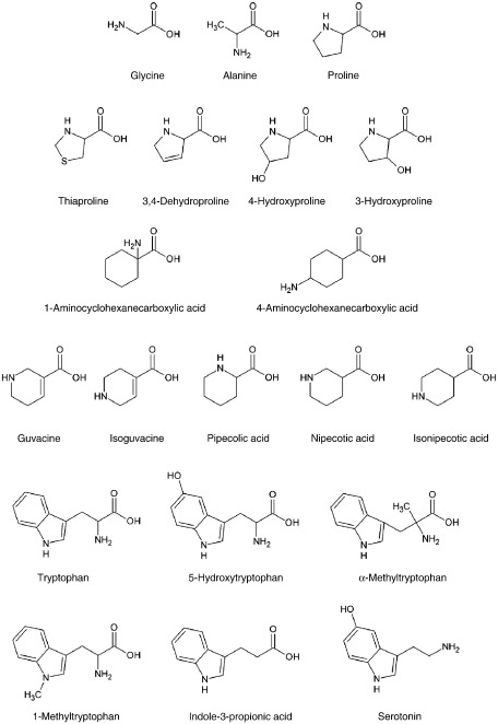
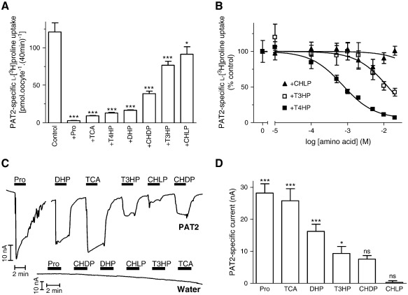
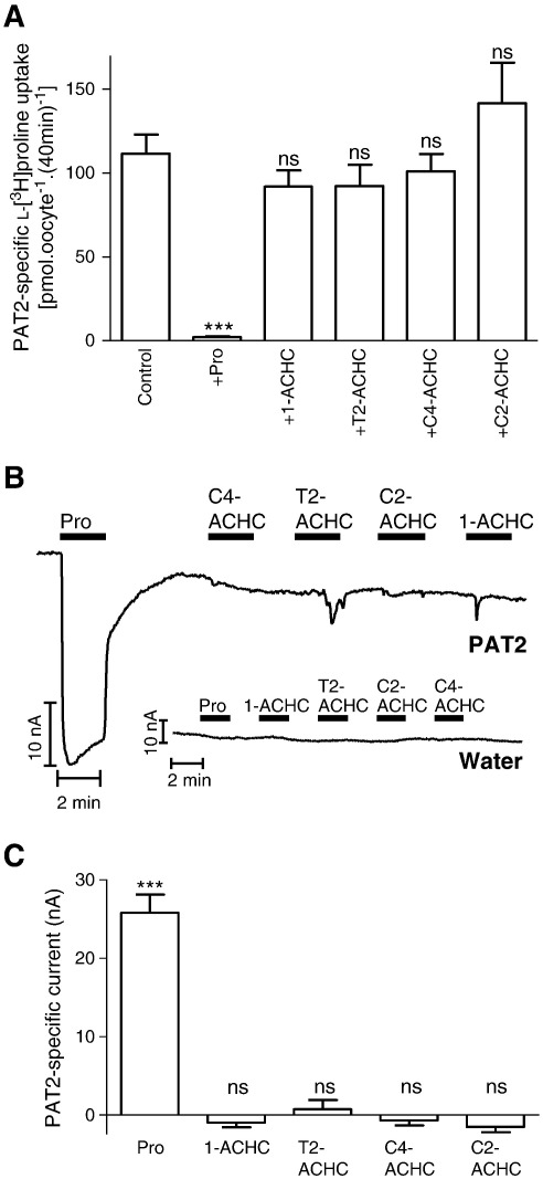
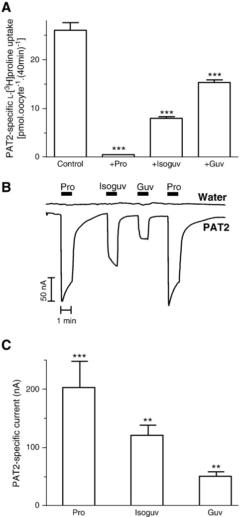
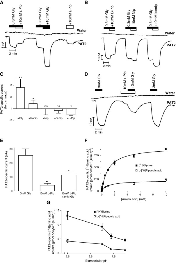
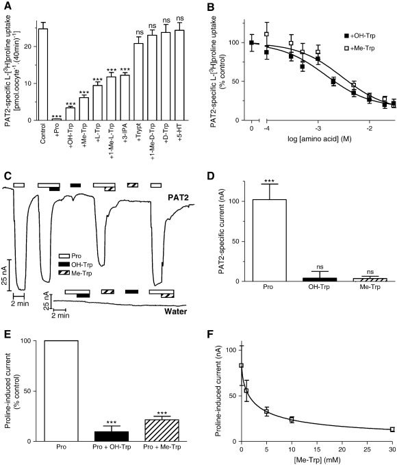
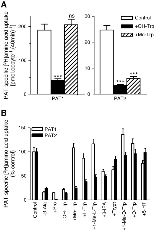
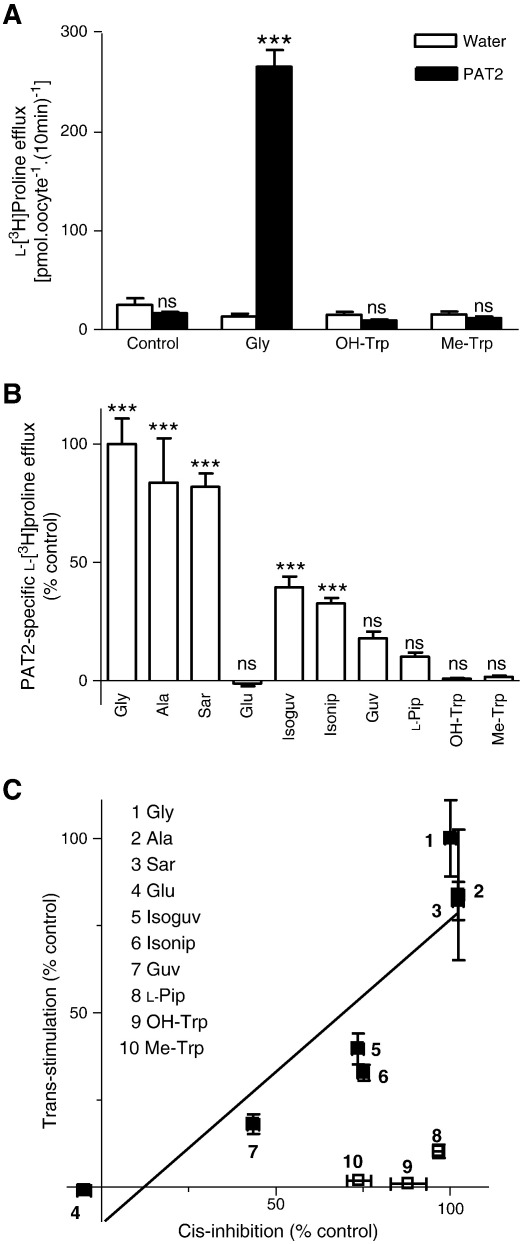
The H(+)-coupled amino acid transporter PAT2 (SLC36A2) transports the amino acids proline, glycine, alanine and hydroxyproline. A physiological role played by PAT2 in amino acid reabsorption in the renal proximal tubule is demonstrated by mutations in SLC36A2 that lead to an iminoglycinuric phenotype (imino acid and glycine uria) in humans. A number of proline, GABA and tryptophan derivatives were examined to determine if they function either as transported substrates or non-transported inhibitors of PAT2. The compounds were investigated following heterologous expression of rat PAT2 in Xenopus laevis oocytes. PAT2 function was characterised by: radiotracer uptake and competition (cis-inhibition) studies; radiotracer efflux and trans-stimulation; and measurement of substrate-induced positive inward current by two-electrode voltage-clamp. In general, the proline derivatives appeared to be transported substrates and the relative ability to induce current flow was closely related to the inhibitory effects on PAT2-mediated l-[(3)H]proline uptake. In contrast, certain heterocyclic GABA derivatives (e.g. l-pipecolic acid) were translocated only slowly. Finally, the tryptophan derivatives inhibited PAT2 function but did not undergo transport. l-Proline uptake was inhibited by 5-hydroxy-l-tryptophan (IC(50) 1.6±0.4mM), α-methyl-d,l-tryptophan (3.5±1.5mM), l-tryptophan, 1-methyl-l-tryptophan and indole-3-propionic acid. Although neither 5-hydroxy-l-tryptophan nor α-methyl-d,l-tryptophan were able to elicit inward current in PAT2-expressing oocytes both reduced the current evoked by l-proline. 5-Hydroxy-l-tryptophan and α-methyl-d,l-tryptophan were unable to trans-stimulate l-proline efflux from PAT2-expressing oocytes, confirming that the two compounds act as non-transported blockers of PAT2. These two tryptophan derivatives should prove valuable experimental tools in future investigations of the physiological roles of PAT2.


Figures

Fig. 1
Fig. 1
Structures of amino acids and derivatives (see PubChem website at http://pubchem.ncbi.nlm.nih.gov).
Fig. 2
Fig. 2
Interaction of proline derivatives with the amino acid transporter PAT2. A. l-[3H]Proline (100 μM) uptake (pH 5.5, Na+-free, 40 min) in PAT2-expressing oocytes in the absence (control) and presence of unlabelled proline (Pro) or related derivatives (all 10 mM), l-thiaproline (TCA), trans-4-hydroxy-l-proline (T4HP), 3,4-dehydro-d,l-proline (DHP), cis-4-hydroxy-d-proline (CHDP), trans-3-hydroxy-l-proline (T3HP) and cis-4-hydroxy-l-proline (CHLP). Results are expressed as PAT2-specific uptake (following subtraction of uptake in water-injected oocytes under identical experimental conditions). Data are mean ± SEM (n = 28–30). ***, p < 0.001; *, p < 0.05, versus control. B. Concentration-dependent inhibition of PAT2-mediated l-[3H]proline uptake (as in A above) by trans-4-hydroxy-l-proline (T4HP, filled squares, n = 20), trans-3-hydroxy-l-proline (T3HP, open squares, n = 7), and cis-4-hydroxy-l-proline (CHLP, filled triangles, n = 19) (all 0.01–20 mM). Results are expressed as a percentage of the uptake in the absence of inhibitor (after subtraction of uptake in water-injected oocytes). Data are mean ± SEM. C. Representative current traces from PAT2-expressing and water-injected oocytes upon exposure to proline and related derivatives (all 10 mM) measured by TEVC. D. Mean data for part C showing PAT2-mediated current (following subtraction of current induced in water-injected oocytes) evoked by proline and related derivatives. Data are mean ± SEM (n = 4–5). ***, p < 0.001; *, p < 0.05; ns, p > 0.05, versus current change in water-injected oocytes.
Fig. 3
Fig. 3
Aminocyclohexanecarboxylic acids (ACHCs) do not interact with PAT2. A. l-[3H]Proline (100 μM) uptake (pH 5.5, Na+-free) was measured in the absence (control) and presence of proline (Pro) and various ACHCs (all 10 mM), 1-aminocyclohexanecarboxylic acid (1-ACHC), trans-2-aminocyclohexanecarboxylic acid (T2-ACHC), cis-4-aminocyclohexanecarboxylic acid (C4-ACHC) and cis-2-aminocyclohexanecarboxylic acid (C2-ACHC). Data are mean ± SEM (n = 18–20). ***, p < 0.001; ns, p > 0.05, versus control. B. Representative current traces observed using PAT2 cRNA- or water-injected oocytes during exposure to proline (Pro) and various aminocyclohexanecarboxylic acids (all 10 mM). C. Mean data for part B showing PAT2-specific current change. Data are mean ± SEM (n = 4). ***, p < 0.001; ns, p > 0.05, versus current change in water-injected oocytes.
Fig. 4
Fig. 4
Interaction of guvacine and isoguvacine with PAT2. A. l-[3H]Proline (10 μM) uptake (pH 5.5, Na+-free) was measured in the absence (control) and presence of proline (Pro), isoguvacine (Isoguv) and guvacine (Guv) (all 10 mM). Data are mean ± SEM (n = 20). ***, p < 0.001, versus control. B. Representative current trace observed using PAT2-expressing and water-injected oocytes during exposure to proline (Pro), isoguvacine (Isoguv) and guvacine (Guv) (all 10 mM). C. Mean data for part B showing PAT2-specific currents. Data are mean ± SEM (n = 4). ***, p < 0.001; **, p < 0.01, versus water-injected oocytes.
Fig. 5
Fig. 5
Interaction of piperidine carboxylates with PAT2. Representative current traces using PAT2-expressing and water-injected oocytes during exposure to glycine (Gly, 0.3 or 10 mM) and/or: A. l-pipecolic acid (l-Pip); B. d-pipecolic acid (d-Pip), nipecotic acid (Nip), isonipecotic acid (Isonip) (all 10 mM). C. Mean PAT2-specific current observed in parts A–B. Data represent the relative change in current observed when glycine (+ Gly), isonipecotic acid (+ Isonip), nipecotic acid (+ Nip), d-pipecolic acid (+ d-Pip) or l-pipecolic acid (+ l-Pip) (all 10 mM) are added to the superfusate in the continued presence of 0.3 mM glycine (control). Results are expressed as the fold change in current relative to that caused by 0.3 mM glycine. Data for glycine (n = 10) and l-pipecolic acid (n = 5) are mean ± SEM, and for isonipecotic acid, nipecotic acid and d-pipecolic acid are mean ± SD (n = 3). **, p < 0.01; *, p < 0.05; ns, p > 0.05, versus the current observed in the presence of 0.3 mM glycine (paired, two-tailed Student's t-test). D. Representative current traces using PAT2-expressing or water-injected oocytes during exposure to a saturating concentration (3 mM) of glycine (Gly) in the presence and absence of 10 mM l-pipecolic acid (l-Pip). E. Mean data for part D showing PAT2-specific current. Data are mean ± SEM (n = 4). **, p < 0.01; *, p < 0.05, versus the current observed in the presence of 3 mM glycine alone. F. [3H]Glycine (filled squares) and l-[3H]pipecolic acid (open squares) (both 0.01–10 mM) uptake (pH 5.5, Na+-free) in PAT2-expressing oocytes. Results are expressed as PAT2-specific uptake (following subtraction of uptake in water-injected oocytes). Data are mean ± SEM (n = 10). G. pH-Dependent [3H]glycine (filled squares) and l-[3H]pipecolic acid (open squares) (both 10 μM) uptake in PAT2-expressing oocytes at extracellular pH 5.5–8.0 (Na+-free). Results are expressed as PAT2-specific uptake. Data are mean ± SEM (n = 9–10).
Fig. 6
Fig. 6
Interaction of tryptophan and related derivatives with PAT2. A. l-[3H]Proline (10 μM) uptake (pH 5.5, Na+-free) in PAT2-expressing oocytes in the absence (control) and presence of proline (Pro) or tryptophan and related derivatives (all 10 mM), 5-hydroxy-l-tryptophan (OH-Trp), α-methyl-d,l-tryptophan (Me-Trp), l-tryptophan (l-Trp), 1-methyl-l-tryptophan (1-Me-l-Trp), indole-3-propionic acid (3-IPA), tryptamine (Trypt), 1-methyl-d-tryptophan (1-Me-d-Trp), d-tryptophan (d-Trp) and serotonin (5-HT). Results are expressed as PAT2-specific uptake. Data are mean ± SEM (n = 19–20). ***, p < 0.001; ns, p > 0.05, versus control. B. l-[3H]Proline (10 μM) uptake (pH 5.5, Na+-free) in the absence and presence of 5-hydroxy-l-tryptophan (OH-Trp) or α-methyl-d,l-tryptophan (Me-Trp) (both 0.1–30 mM). Results are expressed as a percentage of the uptake in the absence of inhibitor (control). Data are mean ± SEM (n = 19–20). C. Representative current traces in PAT2-expressing or water-injected oocytes (pH 5.5, Na+-free) during exposure to 0.2 mM proline (Pro, open bar) in the presence or absence of 20 mM OH-Trp (solid bar) and 20 mM Me-Trp (hatched bar). D. Mean PAT2-specific current evoked by proline (0.2 mM, open column), OH-Trp (filled column) or Me-Trp (hatched column) (both 20 mM), as shown in part C. Data are mean ± SEM (n = 5–6). ***, p < 0.001; ns, p > 0.05, versus water-injected oocytes. E. Mean PAT2-mediated current (in part C) when oocytes are exposed to proline (0.2 mM) in the absence (Pro, open column) or presence of either OH-Trp (Pro + OH-Trp, filled column) or Me-Trp (Pro + Me-Trp, hatched column) (both 20 mM). Results are expressed as a percentage of the control (the current observed in the presence of 0.2 mM proline alone). ***, p < 0.001 versus control. Data are mean ± SEM (n = 5–6). F. Concentration-dependent inhibition by Me-Trp (1–30 mM) of PAT2-mediated proline-induced current (0.2 mM). Data are mean ± SEM (n = 5).
Fig. 7
Fig. 7
Inhibition of PAT1- and PAT2-mediated amino acid uptake by tryptophan derivatives. A. [3H]β-Alanine (100 μM, for PAT1) and l-[3H]proline (10 μM, for PAT2) uptake were measured (pH 5.5, Na+-free) in the absence (control, open columns) and presence of 5-hydroxy-l-tryptophan (OH-Trp, filled columns) or α-methyl-d,l-tryptophan (Me-Trp, hatched columns) (both 10 mM). Data are mean ± SEM (n = 20). ***, p < 0.001; ns, p > 0.05, versus control. B. PAT1 (open columns) and PAT2 (filled columns) mediated amino acid uptake (conditions as in A) in the absence (control) and presence of β-alanine (β-Ala), proline (Pro), 5-hydroxy-l-tryptophan (OH-Trp), α-methyl-d,l-tryptophan (Me-Trp), l-tryptophan (l-Trp), 1-methyl-l-tryptophan (1-Me-l-Trp), indole-3-propionic acid (3-IPA), tryptamine (Trypt), 1-methyl-d-tryptophan (1-Me-d-Trp), d-tryptophan (d-Trp) and serotonin (5-HT) (all 10 mM). Data are mean ± SEM (n = 19–20). Results are expressed as percent control (uptake in absence of inhibitor).
Fig. 8
Fig. 8
Trans-stimulation of PAT2-mediated l-[3H]proline efflux from oocytes discriminates between PAT2 substrates and non-transported inhibitors. PAT2-expressing and water-injected oocytes pre-loaded with l-[3H]proline (approximately 4 mM) were incubated (10 min) in pH 5.5 Na+-free solution in the presence or absence of various amino acids (all 10 mM). A. A typical trans-stimulation experiment to illustrate efflux of l-[3H]proline from PAT2-expressing (filled columns) and water-injected (open columns) oocytes in the absence (control) or presence of glycine (Gly), 5-hydroxy-l-tryptophan (OH-Trp) or α-methyl-d,l-tryptophan (Me-Trp) (all 10 mM). Data are mean ± SEM (n = 10). ***, p < 0.001; ns, p > 0.05, versus water-injected oocytes. B. l-[3H]Proline efflux was measured in the absence (control) or presence of glycine (Gly), l-alanine (Ala), sarcosine (Sar), glutamic acid (Glu), isoguvacine (Isoguv), isonipecotic acid (Isonip), guvacine (Guv), l-pipecolic acid (l-Pip), 5-hydroxy-l-tryptophan (OH-Trp) and α-methyl-d,l-tryptophan (Me-Trp) (all 10 mM). Results are expressed as the percentage of l-[3H]proline efflux in the presence of 10 mM extracellular glycine. Data are mean ± SEM (n = 5–10). ***, p < 0.001; ns, p > 0.05, versus efflux under control conditions (efflux from water-injected oocytes under identical conditions). C. Relationship between the abilities of various amino acids and derivatives to cis-inhibit PAT2-mediated amino acid uptake and trans-stimulate PAT2-mediated amino acid efflux. For cis-inhibition, l-[3H]proline (10 μM) uptake (pH 5.5, Na+-free) was measured in PAT2-expressing oocytes in the absence and presence of unlabelled amino acids (all 10 mM). Results were determined as PAT2-specific uptake (following subtraction of uptake in water-injected oocytes under identical experimental conditions) and are expressed as a percent of the inhibition observed in the presence of 10 mM glycine. Data are mean ± SEM (n = 20). For trans-stimulation, data are taken from panel B, and are expressed as the percent of the trans-stimulation observed in the presence of 10 mM glycine. The relationship [excluding l-pipecolic acid (l-Pip), 5-hydroxy-l-tryptophan (OH-Trp) and α-methyl-d,l-tryptophan (Me-Trp)] was analysed by linear regression (r2 = 0.82).

References

    1. Chen Z., Kennedy D.J., Wake K.A., Zhuang L., Ganapathy V., Thwaites D.T. Structure, tissue expression pattern, and function of the amino acid transporter rat PAT2. Biochem. Biophys. Res. Commun. 2003;304:747–754. - PubMed
    1. Boll M., Foltz M., Rubio-Aliaga I., Daniel H. A cluster of proton/amino acid transporter genes in the human and mouse genomes. Genomics. 2003;82:47–56. - PubMed
    1. Bermingham J.R., Pennington J. Organization and expression of the SLC36 cluster of amino acid transporter genes. Mamm. Genome. 2004;14:114–125. - PubMed
    1. Hediger M.A., Romero M.F., Peng J.B., Rolfs A., Takanaga H., Bruford E.A. The ABCs of solute carriers: physiological, pathological and therapeutic implications of human membrane transport proteins. Pflügers Arch. 2004;447:465–468. - PubMed
    1. Fredriksson R., Nordström K.J.V., Stephansson O., Hägglund M.G.A., Schiöth HB H.B. The solute carrier (SLC) complement of the human genome: phylogenetic classification reveals four major families. FEBS Lett. 2008;582:3811–3816. - PubMed

Publication types