Review
. 2010 Aug 30;62(11):1064-79.
doi: 10.1016/j.addr.2010.07.009. Epub 2010 Aug 4.

Nanoparticle-based theranostic agents

Affiliations
Review

Nanoparticle-based theranostic agents

Jin Xie et al. Adv Drug Deliv Rev. .

Abstract

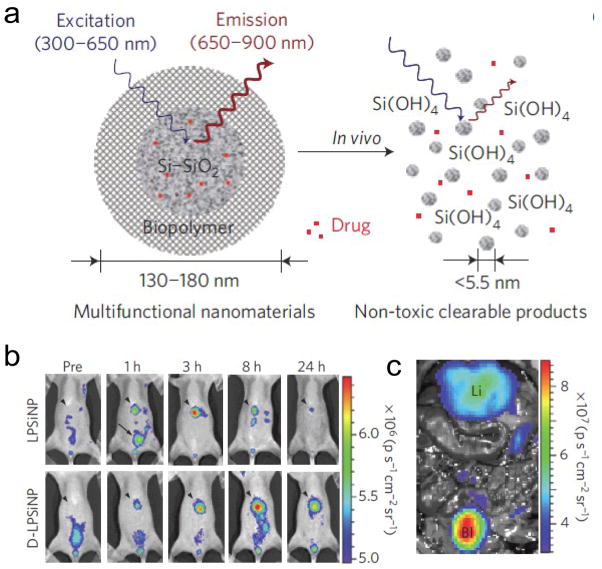
Theranostic nanomedicine is emerging as a promising therapeutic paradigm. It takes advantage of the high capacity of nanoplatforms to ferry cargo and loads onto them both imaging and therapeutic functions. The resulting nanosystems, capable of diagnosis, drug delivery and monitoring of therapeutic response, are expected to play a significant role in the dawning era of personalized medicine, and much research effort has been devoted toward that goal. A convenience in constructing such function-integrated agents is that many nanoplatforms are already, themselves, imaging agents. Their well-developed surface chemistry makes it easy to load them with pharmaceutics and promote them to be theranostic nanosystems. Iron oxide nanoparticles, quantum dots, carbon nanotubes, gold nanoparticles and silica nanoparticles, have been previously well investigated in the imaging setting and are candidate nanoplatforms for building up nanoparticle-based theranostics. In the current article, we will outline the progress along this line, organized by the category of the core materials. We will focus on construction strategies and will discuss the challenges and opportunities associated with this emerging technology.


Figures

Fig. 1
Fig. 1
(a) Illustrative demonstration of the formation of the siRNA-IONP nanoconjugate, which consists of magnetic nanoparticles labeled with near-infrared (NIRF) dye Cy5.5 and coupled with membrane translocation peptides (MPAP), and siRNA targeting GFP (switched to Survivin targeted siRNA in c, d, e). (b) In vivo NIRF optical imaging of mice bearing bilateral 9L-GFP and 9L-RFP tumors 48 h after intravenous probe injection. There was a marked decrease in 9L-GFP-associated fluorescence and no change in 9L-RFP fluorescence. (c-e) Ex vivo assays on tumor samples after siSurvivin-IONP conjugate treatment. (c) Quantitative RT-PCR analysis of survivin expression in LS174T tumors after mice were injected with either MN-NIRF-siSurvivin, a mismatch control, or the parent magnetic nanoparticle alone (MN). (d) Areas of high density apoptotic nuclei (green) were found in tumors treated with MN-NIRF-siSurvivin (left), but not in controls that received only parent magnetic nanoparticles (right). (e) H&E staining of frozen tumor sections revealed considerable eosinophilic areas of tumor necrosis (N) in tumors treated with MN-NIRF-siSurvivin (left). Tumors treated with magnetic nanoparticles were devoid of necrotic tissue (right). Purple hematoxiphilic regions (V) indicate viable tumor tissues. Scale bar, 50μm. Reprinted with permission from ref [44].
Fig. 2
Fig. 2
(a) Schematic illustration of the preparation (upper) and assembly (middle) of LipoMag and reverse-phase evaporated magnetic liposomes (lower). (b-c) MKN-74- and NUGC-4-innoculated mice were given LipoMag-siRNA at 0, 2, 4, 6, 8, 10, 12 and 14 days after the initiation of treatment. The treatment schedule was as follows: Group A = control. Group B, C, D were injected with LipoMag loaded with control siRNA sequence. And among them, B was treated without magnetic field. C and D were treated with internal and external magnetic fields, respectively. Group E, F, G were given LipoMag loaded with the modified siRNA-EGFR#4 sequence. E was given the therapeutics without magnetic filed. F and G were injected with nanoparticle therapeutics with internal and external magnetic fields, respectively. (b) Tumor growth curve for each treatment over time. Only the LipoMag/siRNAEGFR groups under a magnetic field showed a significant anti-tumor effect. (c) Two days after the last treatment, the degree of angiogenesis, proliferation and apoptosis were assayed by immunostaining of vWF, Ki-67 and ssDNA, respectively. Reprinted with permission from ref [50].
Fig. 3
Fig. 3
(a) & (b) Formation and working mechanism of QD-Apt(Dox)-FRET nanosystem. (a) CdSe/ZnS QDs are surface functionalized with the A10 PSMA aptamer. The intercalation of Dox within the A10 PSMA aptamer on the surface of QDs resulted in the formation of the QD-Apt(Dox), which quenched fluorescence from both QD and Dox (“OFF” state). (b) Schematic illustration of specific uptake of QD-Apt(Dox) conjugates into target cancer cell through PSMA mediate endocytosis. The release of Dox from the QD-Apt(Dox) conjugates induced the recovery of fluorescence from both QD and Dox (“ON” state), thereby sensing the intracellular delivery of Dox and enabling the synchronous fluorescent localization and killing of cancer cells. (c) & (d) Confocal laser scanning microscopy images of PSMA-expressing LNCaP cells after incubation with 100 nM QD-Apt(Dox) conjugates for 0.5 h at 37 °C, washing two times with PBS buffer, and further incubation at 37 °C for (c) 0 h and (d) 1.5 h. Dox and QD are shown in red and green, respectively, and the lower right images of each panel represent the overlay of Dox and QD fluorescence. Reprinted with permission from ref [76].
Fig. 4
Fig. 4
(a) Schematic illustration of gold nanoshell synthesis and bioconjugation. (b-d) In vivo photothermal ablation with targeted NDP-MSH-PEG-HAuNS to induce selective destruction of B16/F10 melanoma in nude mice. (b) [18F] fluorodeoxyglucose PET imaging showed significantly reduced metabolic activity in tumors after photothermal ablation in mice pretreated with NDP-MSH-PEG-HAuNS, but not in mice pretreated with PEG-HAuNS or saline. T, tumor. Arrowheads, tumors irradiated with near-IR light. (c) [18F] fluorodeoxyglucose uptake (%ID/g) before and after laser treatment. (d) Histologic assessment of tumor necrosis with H&E staining. Reprinted with permission from ref [122].
Fig. 5
Fig. 5
(a) Scheme for the conjugation and photothermal-activated release of siRNA. (b) Postulated uptake and siRNA release mechanisms of the folated, siRNA-Au shell conjugates. (c) Photothermal-induced endolysosomal escape of Dy547-labeled siRNA. Green, LysoTracker Green-labeled endolysosomes; red, Dy547-labeled siRNA. Scale bar, 10 μm. (d) & (e), effect of p65 siRNA photothermal transfection combined with irinotecan on nude mice bearing HeLa cancer xenografts. (c) Representative micrographic images of tumors stained with H&E. (d) Tumor size versus time curve. Control, tumor-bearing mice did not receive any treatment. Reprinted with permission from ref [123].
Fig. 6
Fig. 6
(a) Schematic illustration of the PTX conjugation to SWNT functionalized by phospholipids with branched PEG chains. (b) Tumor growth curves of 4T1 tumor-bearing mice that received different treatments. (c) Scheme for DOX-SWNT complex formation. (d) Tumor growth curves. Raji-tumor-bearing SCID mice were treated with different DOX formulations. Reprinted with permission from ref [156] and [158].
Fig. 7
Fig. 7
(a) Schematic diagram depicting the structure and in vivo degradation process of the silica nanoparticles. (b) In vivo images of luminescent porous silicon nanoparticles (LPSiNPs) and dextran coated LPSiNPs (D-LPSiNPs). The mice were imaged at multiple time points after intravenous injection of LPSiNPs and D-LPSiNPs (20 mg/kg). Arrowheads and arrows with solid lines indicate liver and bladder, respectively. (c) In vivo image showing the clearance of a portion of the injected dose of LPSiNPs into the bladder, 1 h post-injection. Li and Bl indicate liver and bladder, respectively. Reprinted with permission from ref [188].

Similar articles

Cited by

References

    1. Del Vecchio S, Zannetti A, Fonti R, Pace L, Salvatore M. Nuclear imaging in cancer theranostics. Q J Nucl Med Mol Imaging. 2007;51:152–163. - PubMed
    1. Nie S, Xing Y, Kim GJ, Simons JW. Nanotechnology applications in cancer. Annu Rev Biomed Eng. 2007;9:257–288. - PubMed
    1. Liu Y, Miyoshi H, Nakamura M. Nanomedicine for drug delivery and imaging: a promising avenue for cancer therapy and diagnosis using targeted functional nanoparticles. Int J Cancer. 2007;120:2527–2537. - PubMed
    1. Cai W, Chen X. Multimodality molecular imaging of tumor angiogenesis. J Nucl Med. 2008;49(Suppl 2):113S–128S. - PubMed
    1. Cai W, Chen X. Nanoplatforms for targeted molecular imaging in living subjects. Small. 2007;3:1840–1854. - PubMed

Publication types