. 2010 Aug 13;87(2):265-73.
doi: 10.1016/j.ajhg.2010.07.003.

Mutations in PVRL4, encoding cell adhesion molecule nectin-4, cause ectodermal dysplasia-syndactyly syndrome

Affiliations

Mutations in PVRL4, encoding cell adhesion molecule nectin-4, cause ectodermal dysplasia-syndactyly syndrome

Francesco Brancati et al. Am J Hum Genet. .

Abstract

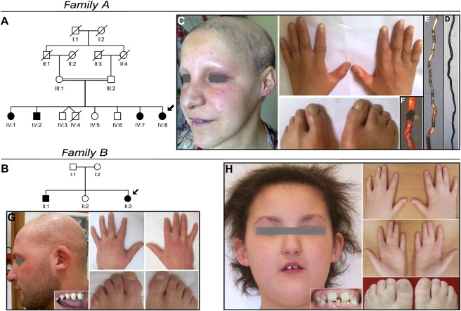
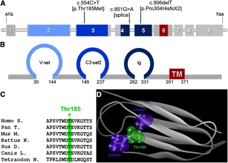
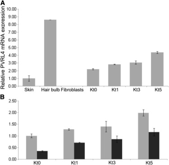
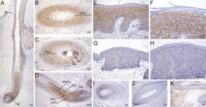
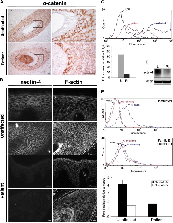
Ectodermal dysplasias form a large disease family with more than 200 members. The combination of hair and tooth abnormalities, alopecia, and cutaneous syndactyly is characteristic of ectodermal dysplasia-syndactyly syndrome (EDSS). We used a homozygosity mapping approach to map the EDSS locus to 1q23 in a consanguineous Algerian family. By candidate gene analysis, we identified a homozygous mutation in the PVRL4 gene that not only evoked an amino acid change but also led to exon skipping. In an Italian family with two siblings affected by EDSS, we further detected a missense and a frameshift mutation. PVRL4 encodes for nectin-4, a cell adhesion molecule mainly implicated in the formation of cadherin-based adherens junctions. We demonstrated high nectin-4 expression in hair follicle structures, as well as in the separating digits of murine embryos, the tissues mainly affected by the EDSS phenotype. In patient keratinocytes, mutated nectin-4 lost its capability to bind nectin-1. Additionally, in discrete structures of the hair follicle, we found alterations of the membrane localization of nectin-afadin and cadherin-catenin complexes, which are essential for adherens junction formation, and we found reorganization of actin cytoskeleton. Together with cleft lip and/or palate ectodermal dysplasia (CLPED1, or Zlotogora-Ogur syndrome) due to an impaired function of nectin-1, EDSS is the second known "nectinopathy" caused by mutations in a nectin adhesion molecule.


Figures

Figure 1
Figure 1
Pedigrees and Clinical Manifestations of Two Families with EDSS (A and B) Pedigree structure of families A and B. The probands are indicated by arrows, clear symbols represent unaffected individuals, and filled symbols represent affected individuals. (C) Patient IV:8, aged 25 years, shows diffuse alopecia of the scalp and absent eyebrows and eyelashes. Partial skin syndactyly of fingers 2-3 and toes 2-3 and 4-5 is present bilaterally. (D–F) Hair shaft abnormalities in this patient observed at light microscopy (D) consist of peculiar repeated twists of the hair (pili torti), whereas polarized microscopy (E) outlines high-frequency bands with consequent alteration of the normal banding pattern of the hair shaft (magnification 40×). Trichoschisis with transverse fracture is evident with polarized microscopy magnification 100× (F). (G and H) Family B, affected siblings aged 27 and 9 years. The older brother (G) shows alopecia, widely spaced teeth with conical crowns, and partial cutaneous syndactyly of toes 2-3. Fingers 2-3 and 3-4 syndactyly has been surgically corrected. The proband (H) shows short and coarse uncombable hair, sparse eyebrows and eyelashes, widely spaced irregular teeth, and syndactyly of fingers 2-3-4 and of toes 2-3 and 4-5.
Figure 2
Figure 2
Mutations Identified in the PVRL4 Gene (A) Schematic view of the human PVRL4 gene and localization of identified mutations. (B) Schematic representation of nectin-4 exhibiting three extracellular Ig-like domains (blue) and a transmembrane domain (red). The numbers denote the amino acids located at the boundaries of each domain. (C) Amino acid sequence alignment showing conservation among species of the mutated Thr185 residue (). (D) Modeled structure of nectin-4 in the amino acid range 147–244 encompassing the second Ig-like domain. Site of mutation Thr185 (green cloud) and its interacting amino acids Val187 and Leu221 (blue clouds) are shown.
Figure 3
Figure 3
Nectin-4 mRNA Expression in Specimens from Unaffected Individual and EDSS Patient (A) Relative PVRL4 mRNA expression, as determined by quantitative PCR in skin biopsy, plucked hair bulbs, primary fibroblasts, and proliferating (Kt0) and differentiated (Kt1, Kt3, Kt5) keratinocytes obtained from healthy donors. Keratinocyte differentiation was induced by the addition of 1.2 mM calcium to the culture media for 1 (t1), 3 (t3), or 5 (t5) days. PVRL4 is expressed in skin, keratinocytes, and mainly in hair bulbs, but not in fibroblasts. Expression level increases during keratinocyte differentiation. (B) Analysis of PVRL4 expression in primary keratinocytes at different stages (Kt0–Kt5) from family B patient II:1 (black column) and from a control individual (gray column). Note the strongly reduced expression in the patient's samples. Experiments were run in duplicate. Error bars are ± standard deviation. The relative expression values were determined via the ΔΔCt method.
Figure 4
Figure 4
Localization of Nectin-4 in Human Skin and Hair (A–F) Frozen sections of human skin containing terminal hair obtained from healthy donors were stained with the mouse anti-nectin-4 monoclonal antibody (N4.61), as previously described. In the hair follicle (A–D), a pericellular signal is first detected at the level of the hair bulb in suprabasal matrix epithelial cells (M), giving rise to the different specialized hair follicle compartments. The hair shaft cortex and cuticle (HSC and HSCu) (D), as well as all the inner root sheath (IRS) layers (i.e., Henle's [He], Huxley's [Hu], and cuticle [IRSCu]) (C and D) appear strongly positive until the onset of keratinization, which occurs at first in the Henle's layer, then in the IRS and hair shaft cuticle, and finally in the Huxley's layer and hair shaft cortex (A, C, and D). A less intense staining is present in all suprabasal layers of the outer root sheath (ORS) (A–D). The epidermis is also stained, and the signal intensity shows an increasing gradient from the first suprabasal to the granular layer; the signal disappears at the transition zone between the granular layer and stratum corneum (E and F). (G–J) Nectin-4 staining is strongly reduced in the epidermis of family B patient II:1 (G and H) and in his hair follicles (I and J). (K) Nonspecific labeling is observed to be limited to the sebaceous glands with the secondary biotin-conjugated antibody alone. Scale bars represent 100 μm (A, D, and I–K), 25 μm (B and C), and 50 μm (E–H).
Figure 5
Figure 5
Tissue and Cellular Consequences of Nectin-4 Alteration (A) Frozen sections of human skin with terminal hair obtained from an unaffected individual and family B patient II:1 were immunostained with the AJ marker α-catenin (clone 7A4, Invitrogen). Higher-magnification images of the boxed regions are shown. In the control sample, α-catenin preferentially stains along cell-cell adhesion sites in all nonkeratinized structures of the hair. In the patient, although a similar staining is observed along the cell-cell junctions at the IRS level, its distribution is altered in the ORS. Scale bars represent 50 μm. (B) Nectin-4 and F-actin were stained in serial sections of human skin from an unaffected individual and family B patient II:1 with anti-nectin-4 mAb (N4.61) or rhodamine-phalloidin (Invitrogen), respectively. Although strong reduction of nectin-4 expression does not seem to affect the cellular architecture at the interfollicular epidermis level (top), the actin cytoskeleton is strongly disorganized in the hair follicle (bottom). Scale bars represent 25 μm. (C) FACS analysis on dissociated keratinocytes showing a decrease of cell surface expression of nectin-4 in family B patient II:1 (red) compared to a control sample (blue). The relative histogram documents the strong reduction of expression (relative to IgG1) between mean values of three unaffected control samples (U, light gray) and the patient (Pt, dark gray). Cells were incubated with a mouse IgG1 anti-nectin-4 IgV domain monoclonal antibody (N4.61) or an irrelevant mouse IgG1 monoclonal antibody (5 μg/ml). After washes, cells were incubated with a phycoerythrin-labeled goat anti-mouse secondary antibody (Immunotech) (1/100). Incubation conditions were 60 min at +4°C. (D) Immunoblot analysis of nectin-4, performed with monoclonal antibody (BAF2659; R&D Systems) on protein extracts of differentiated primary keratinocytes from an unaffected individual and family B patient II:1, confirming strong reduction of protein expression. (E) Trans-heterophilic interaction between nectin-1 and nectin-4 was determined by FACS analysis by using binding of nectin-1-Fc soluble recombinant protein, as previously described. A nectins trans-interactive network exists with nectin-1 binding nectin-3 and nectin-4 and with nectin-2 binding nectin-3. We found the absence of nectin-3 expression in control keratinocytes (data not shown) and thus used nectin-2-Fc as a negative binding control. The histogram represents nectin-1-Fc (black) and nectin-2-Fc (white) binding expression (relative to control) in keratinocytes from family B patient II:1 compared to three unaffected individuals. In patient's keratinocytes, nectin-1 to nectin-4 binding was dramatically reduced.
Figure 6
Figure 6
Nectin-4 Expression during Mouse Limb Development (A) Relative Pvrl4 mRNA expression in whole-limb mRNA at different embryonic stages determined by quantitative PCR. Three to five limbs were pooled for mRNA isolation. The relative expression values were determined via the ΔΔCt method. Experiments were run in duplicate. Error bars are ± standard deviation. (B) Whole-mount in situ hybridization for Pvrl4 mRNA expression in E15.5 mouse limb denotes strong expression in areas where digit separation is ongoing.

Similar articles

Cited by

References

    1. Fuchs E. Scratching the surface of skin development. Nature. 2007;445:834–842. - PMC - PubMed
    1. Gumbiner B.M. Cell adhesion: The molecular basis of tissue architecture and morphogenesis. Cell. 1996;84:345–357. - PubMed
    1. Perez-Moreno M., Jamora C., Fuchs E. Sticky business: Orchestrating cellular signals at adherens junctions. Cell. 2003;112:535–548. - PubMed
    1. Freire-Maia N. Ectodermal dysplasias. Hum. Hered. 1971;21:309–312. - PubMed
    1. Freire-Maia N., Lisboa-Costa T., Pagnan N.A. Ectodermal dysplasias: How many? Am. J. Med. Genet. 2001;104:84. - PubMed

Publication types

MeSH terms