

Follicular helper T cell differentiation requires continuous antigen presentation that is independent of unique B cell signaling
- PMID: 20691615
- PMCID: PMC3433066
- DOI: 10.1016/j.immuni.2010.07.015
Follicular helper T cell differentiation requires continuous antigen presentation that is independent of unique B cell signaling
Abstract
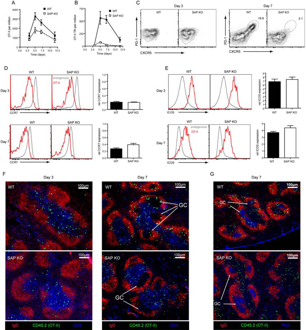
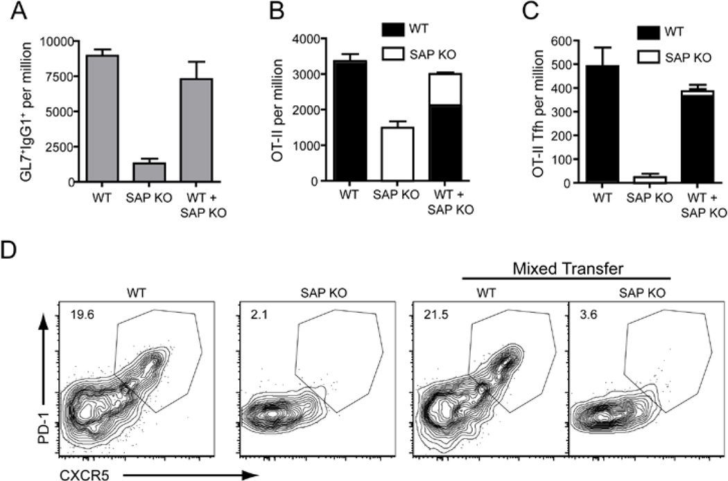
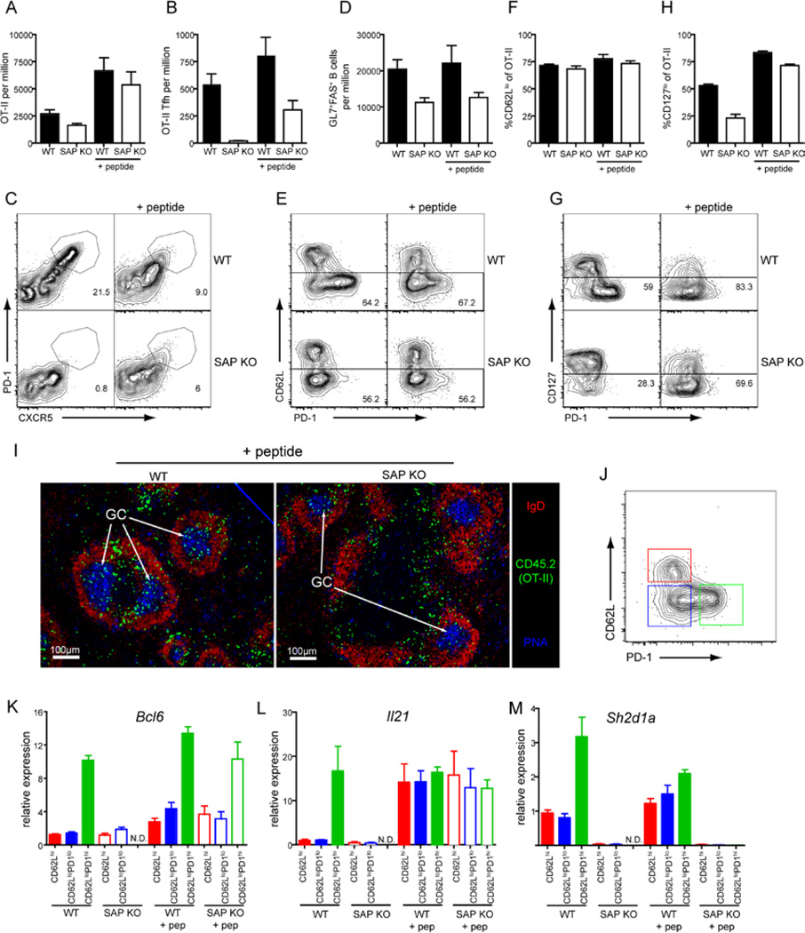
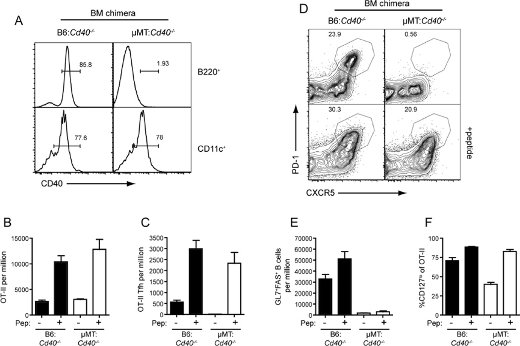
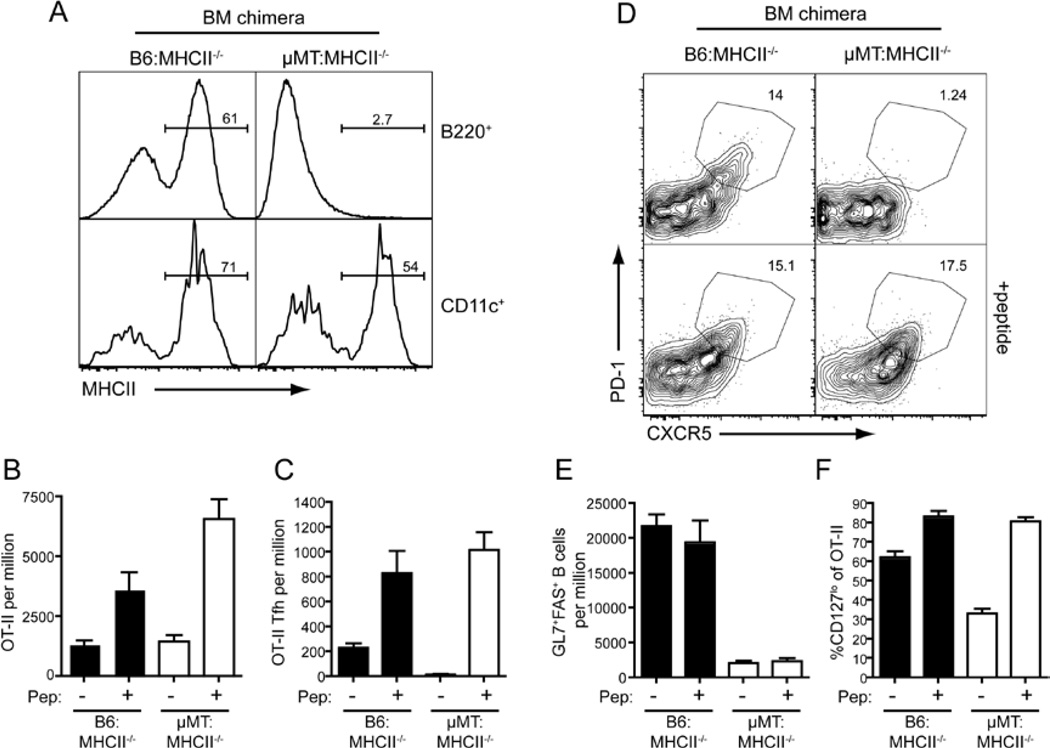
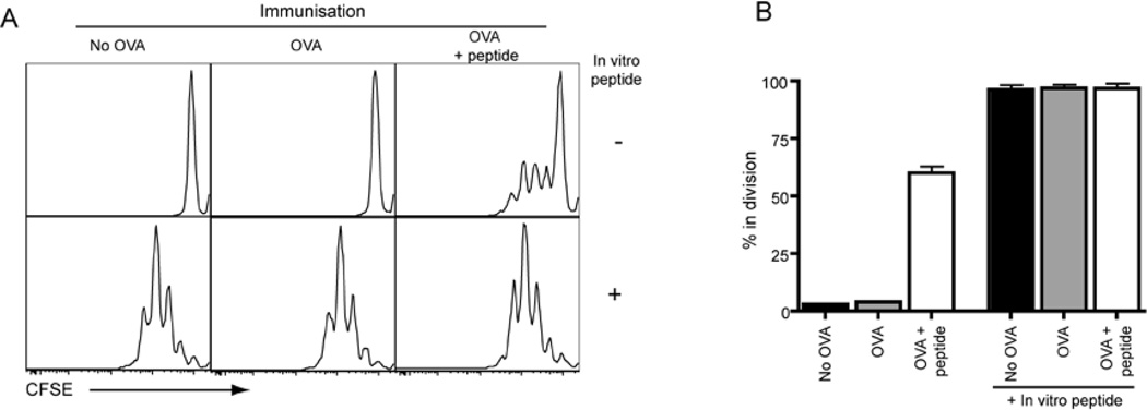
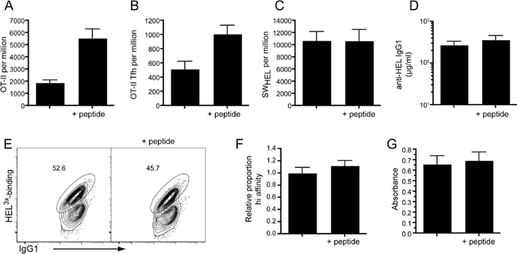
Effective humoral immunity depends on the support of B cell responses by T follicular helper (Tfh) cells. Although it has been proposed that Tfh cell differentiation requires T-B interactions, the relative contribution of specific populations of Ag-presenting cells remains unknown. We employed three independent strategies that compromised interactions between CD4(+) T cells and activated B cells in vivo. Whereas the expansion of CD4(+) T cells was relatively unaffected, Tfh cell differentiation was completely blocked in all scenarios. Surprisingly, augmenting antigen presentation by non-B cells rescued Tfh cell differentiation, as determined by surface phenotype, gene expression, and germinal center localization. We conclude that although Ag presentation by responding B cells is typically required for the generation of Tfh cells, this does not result from the provision of a unique B cell-derived signal, but rather because responding B cells rapidly become the primary source of antigen.
Copyright 2010 Elsevier Inc. All rights reserved.
Figures
Similar articles
-
Repression of miR-31 by BCL6 stabilizes the helper function of human follicular helper T cells.Proc Natl Acad Sci U S A. 2017 Nov 28;114(48):12797-12802. doi: 10.1073/pnas.1705364114. Epub 2017 Nov 13. Proc Natl Acad Sci U S A. 2017. PMID: 29133396 Free PMC article.
-
CD25(+) Bcl6(low) T follicular helper cells provide help to maturing B cells in germinal centers of human tonsil.Eur J Immunol. 2015 Jan;45(1):298-308. doi: 10.1002/eji.201444911. Epub 2014 Oct 27. Eur J Immunol. 2015. PMID: 25263533 Free PMC article.
-
Functional and epigenetic studies reveal multistep differentiation and plasticity of in vitro-generated and in vivo-derived follicular T helper cells.Immunity. 2011 Oct 28;35(4):622-32. doi: 10.1016/j.immuni.2011.07.015. Epub 2011 Oct 20. Immunity. 2011. PMID: 22018472 Free PMC article.
-
Follicular Helper T Cells.Annu Rev Immunol. 2016 May 20;34:335-68. doi: 10.1146/annurev-immunol-041015-055605. Epub 2016 Feb 22. Annu Rev Immunol. 2016. PMID: 26907215 Review.
-
How T cells earn the follicular rite of passage.Immunity. 2011 Nov 23;35(5):671-80. doi: 10.1016/j.immuni.2011.11.001. Immunity. 2011. PMID: 22118524 Review.
Cited by
-
Tracking early T follicular helper cell differentiation in vivo.Methods Mol Biol. 2015;1291:27-38. doi: 10.1007/978-1-4939-2498-1_3. Methods Mol Biol. 2015. PMID: 25836299 Free PMC article.
-
Single cell analysis of host response to helminth infection reveals the clonal breadth, heterogeneity, and tissue-specific programming of the responding CD4+ T cell repertoire.PLoS Pathog. 2021 Jun 9;17(6):e1009602. doi: 10.1371/journal.ppat.1009602. eCollection 2021 Jun. PLoS Pathog. 2021. PMID: 34106992 Free PMC article.
-
Residual Proviral Reservoirs: A High Risk for HIV Persistence and Driving Forces for Viral Rebound after Analytical Treatment Interruption.Viruses. 2021 Feb 21;13(2):335. doi: 10.3390/v13020335. Viruses. 2021. PMID: 33670027 Free PMC article. Review.
-
Skin dendritic cells induce follicular helper T cells and protective humoral immune responses.J Allergy Clin Immunol. 2015 Nov;136(5):1387-97.e1-7. doi: 10.1016/j.jaci.2015.04.001. Epub 2015 May 9. J Allergy Clin Immunol. 2015. PMID: 25962902 Free PMC article.
-
Cutting edge: Bcl6-interacting corepressor contributes to germinal center T follicular helper cell formation and B cell helper function.J Immunol. 2015 Jun 15;194(12):5604-8. doi: 10.4049/jimmunol.1500201. Epub 2015 May 11. J Immunol. 2015. PMID: 25964495 Free PMC article.
References
-
- Aicher A, Hayden-Ledbetter M, Brady WA, Pezzutto A, Richter G, Magaletti D, Buckwalter S, Ledbetter JA, Clark EA. Characterization of human inducible costimulator ligand expression and function. J Immunol. 2000;164:4689–4696. - PubMed
-
- Akiba H, Takeda K, Kojima Y, Usui Y, Harada N, Yamazaki T, Ma J, Tezuka K, Yagita H, Okumura K. The role of ICOS in the CXCR5+ follicular B helper T cell maintenance in vivo. J Immunol. 2005;175:2340–2348. - PubMed
-
- Armitage RJ, Fanslow WC, Strockbine L, Sato TA, Clifford KN, Macduff BM, Anderson DM, Gimpel SD, Davis-Smith T, Maliszewski CR. Molecular and biological characterization of a murine ligand for CD40. Nature. 1992;357:80–82. - PubMed
-
- Barnden MJ, Allison J, Heath WR, Carbone FR. Defective TCR expression in transgenic mice constructed using cDNA-based alpha- and beta-chain genes under the control of heterologous regulatory elements. Immunol Cell Biol. 1998;76:34–40. - PubMed
Publication types
MeSH terms
Substances
Grants and funding
LinkOut - more resources
Full Text Sources
Molecular Biology Databases
Research Materials
