. 2010 Oct 15;346(2):258-71.
doi: 10.1016/j.ydbio.2010.07.030. Epub 2010 Aug 5.

Dorsal eye selector pannier (pnr) suppresses the eye fate to define dorsal margin of the Drosophila eye

Affiliations

Dorsal eye selector pannier (pnr) suppresses the eye fate to define dorsal margin of the Drosophila eye

Sarah M Oros et al. Dev Biol. .

Abstract

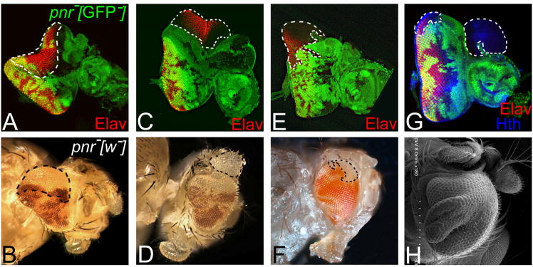
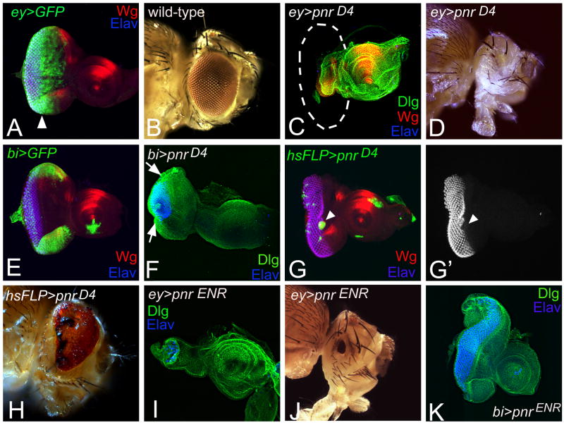
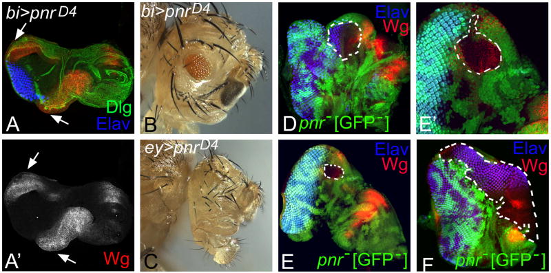
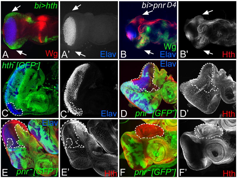
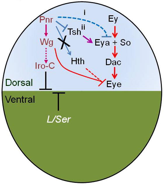
Axial patterning is crucial for organogenesis. During Drosophila eye development, dorso-ventral (DV) axis determination is the first lineage restriction event. The eye primordium begins with a default ventral fate, on which the dorsal eye fate is established by expression of the GATA-1 transcription factor pannier (pnr). Earlier, it was suggested that loss of pnr function induces enlargement in the dorsal eye due to ectopic equator formation. Interestingly, we found that in addition to regulating DV patterning, pnr suppresses the eye fate by downregulating the core retinal determination genes eyes absent (eya), sine oculis (so) and dacshund (dac) to define the dorsal eye margin. We found that pnr acts downstream of Ey and affects the retinal determination pathway by suppressing eya. Further analysis of the "eye suppression" function of pnr revealed that this function is likely mediated through suppression of the homeotic gene teashirt (tsh) and is independent of homothorax (hth), a negative regulator of eye. Pnr expression is restricted to the peripodial membrane on the dorsal eye margin, which gives rise to head structures around the eye, and pnr is not expressed in the eye disc proper that forms the retina. Thus, pnr has dual function, during early developmental stages pnr is involved in axial patterning whereas later it promotes the head specific fate. These studies will help in understanding the developmental regulation of boundary formation of the eye field on the dorsal eye margin.


Figures

Fig. 1
Fig. 1. Pnr expressesion is restricted to the peripodial membrane (PM) of the dorsal eye margin
(A) pnr expression (pnr Gal4 drive UAS-GFP, Singh and Choi, 2003; Singh et al., 2005) is absent in the first instar eye-antennal imaginal disc whereas Wg (red) is expressed in the entire eye disc. Note that pnr expression in the brain at this stage is seen. (B) In the second instar eye-antennal imaginal disc, pnr expression (green) is initiated in 15–20 cells on the dorsal eye margin and Wg (red) is expressed laterally on both dorsal and ventral eye margins. At this stage, Hth (blue) expression is present in the entire eye disc. (C, C′, C″) In the early third instar eye-antennal disc, pnr expression in the dorsal eye margin is restricted only to the peripodial membrane (PM) whereas Hth (blue) is also expressed in peripodial membrane (PM) of the eye-antennal disc. (C′) pnr (green) expression at this stage is absent in the disc proper (DP). Hth (blue) expression begins to retract with the initiation of MF and stays anterior to the furrow (Pai et al., 1998; Bessa et al., 2002; Singh et al., 2002). (C″) Pnr expression is restricted to the peripodial membrane (PM) specific cells on the dorsal eye margin. (D) In the late third instar eye-antennal imaginal disc, pnr (green) expression is restricted to the dorsal eye margin whereas Wg (red) expression is restricted to the dorsal and ventral eye margins. Hth (Blue) is expressed in rings in the proximal region of antenna and expressed both in the dorsal and ventral part of the disc proper anterior to the furrow. Dashed lines indicate the approximate midline, the border between D (Dorsal) and V (ventral) eye. All the eye-antennal imaginal discs and the adult eyes are organized as Dorsal (D) up and the ventral (V) down. Markers for immunostaining are shown in color labels. (AN: Antenna)
Fig. 2
Fig. 2. Loss-of-function of pnr exhibits a range of eye enlargements and antennal duplications in the dorsal eye
(A, B) Loss-of-function clones of pnr in the dorsal eye margin {marked by the absence of GFP (green) in the eye-antennal imaginal disc and absence of the mini-white reporter (red) in the adult eye} results in a non-autonomous ectopic eye enlargement as seen in the eye-antennal imaginal disc and in the adult eye. The ectopic eye enlargements are not restricted within the clone. However, they extend both in the wild-type as well as in the pnr mutant cells of the eye-antennal disc. Note that the dorsal clone boundary is marked by white dotted line in the eye disc and by black dotted line in the adult eye. (C, D) Loss-of-function of pnr in the dorsal eye results in an autonomous ectopic dorsal eye anterior to the normal eye field. These ectopic eyes are restricted to within the clones. Note that not all the cells of the pnr loss-of-function clone differentiate to the photoreceptors. (E, F) Loss-of-function clones of pnr in the dorsal eye have no effect on the eye field as seen in the eye disc and the adult eye. All these clones were restricted to the disc proper. (G, H) Loss-of-function clones of pnr in the antenna results in duplication of the antennal field as seen in (G) the eye-antennal disc and (H) the adult head. (H) Scanning electron microscopy (SEM) of the adult head showing antennal duplication and dorsal eye enlargement (Magnification X180). Note that only a few pnr loss-of-function clones show both dorsal eye enlargements along with the antennal duplication.
Fig. 3
Fig. 3. Pnr suppresses the eye fate
(A) Eye-antennal imaginal disc showing domain of expression of GFP reporter (green) under the ey Gal4 (ey>GFP). Note that the ey Gal4 drives the expression of GFP reporter in the entire eye-antennal imaginal disc (both anterior as well as posterior to the morphogenetic furrow (MF) marked by white arrowhead). (B) Wild-type adult eye. (C, D) Misexpression of pnr in the eye using the ey-Gal4 driver (ey>pnrD4) results in the suppression of eye fate and leads to a “no-eye” phenotype in the (C) eye-antennal disc (Elav, a pan-neural marker, which marks the photoreceptors) as well as in the (D) adult eye. The white dotted line in 3C marks the possible outline of the eye disc. There is no effect on the antennal field both in the eye-antennal imaginal disc as well as the adult head. (E) Another Gal4 driver, bi-Gal4 drives expression of a GFP reporter (bi>GFP) both on the dorsal and the ventral eye disc margin. (F) Misexpression of pnr using bi-Gal4 (bi>pnrD4) results in the suppression of eye fate on both the dorsal and the ventral eye margin as evident from the loss of Elav expression (white arrows). (G, G′, H) Gain-of-function clones of pnr (marked by GFP, white arrowhead) generated by random “flp-out” approach in the eye using the heat shock-FLP showed the suppression of eye as evident from the absence of (G′) Elav in the eye disc as well as (H) in the adult eye. Note that the eye suppression in the pnr heat shock “flp out” clones was seen only in the larger clones. Further, necrosis (black spots) is also seen in the adult eye upon misexpression of pnr in the eye. (I, J) Blocking pnr function in the entire eye using pnr ENR construct (ey>pnrENR) results in a “small eye” phenotype as seen (I) in the eye imaginal disc as well as (J) in the adult eye. (K) However, blocking pnr function both on the dorsal and the ventral eye disc margin (bi>pnr ENR) results in the dorsal eye enlargement whereas there was no effect on the ventral eye margin. This data suggests that pnr suppresses eye on the dorsal eye margin.
Fig. 4
Fig. 4. Pnr suppresses the expression of retinal differentiation genes in the eye
(A, A′, A″) Loss-of-function clones of pnr in the eye exhibit dorsal eye enlargement by (A″) ectopic Elav expression. In these clones where dorsal eye enlargement is seen, (A′) the expression of retinal precursor marker Ey is restricted anterior to the furrow (white arrow). The dorsal eye enlargement, marked by Elav (blue) is the outcome of the pnr loss-of-function clone. Note that Ey is absent in the differentiating photoreceptors. Therefore, Ey is not seen in these clones. (B-D) Loss-of-function clones of pnr showing an ectopic dorsal eye phenotype with ectopic induction of retinal determination genes like (B, B′, B″) Eya (white arrow), (C, C′, C″) So (white arrow), and (D, D′, D″) Dac (white arrow). Note that these retinal determination genes, which act downstream to Ey, and unlike Ey are expressed in the differentiating photoreceptor neurons. (E- H) Gain-of-function of pnr in the eye suppresses the retinal determination genes. (E, E′) Misexpression of pnr on dorsal and ventral eye margins by using a bi-Gal4 driver (bi>pnrD4), results in strong upregulation of Ey on both dorsal and ventral eye margins (marked by a white arrowhead). (F-H) However, misexpression of pnr (bi>pnrD4) suppresses the downstream retinal differentiation genes (F, F′) Eya, (G) So, (H) Dac on both dorsal and ventral margins (marked by arrow heads). The anterior Dac expression (anterior to furrow) went all the way down to the posterior margin in bi>pnrD4 misexpression. Ey marks retinal precursor cells and is required for the specification of eye field. Our results suggest that pnr may not affect early eye specification function of Ey, whereas pnr suppresses the retinal determination genes like eya, so and dac, which acts downstream to Ey.
Fig. 5
Fig. 5. pnr induces downstream target Wg to suppress the eye
Wg is known to act as a negative suppressor of eye fate. Wg is expressed laterally both on the dorsal and the ventral eye margins (Fig. 1B). (A, A′) Misexpression of pnr on both dorsal and ventral eye margin using bi-Gal4 results in the suppression of eye on both DV margins along with ectopic induction of Wg (marked by white arrows). (B) bi>pnrD4 results in the reduction of eye both on the dorsal and ventral eye margins. This phenotype is similar to bi>wg (Singh et al., 2002). (C) Misexpression of Wg in the entire eye using ey-Gal4 (ey>wg) results in “no-eye”. (D) Loss-of-function clones of pnr in the dorsal eye (marked by absence of GFP reporter and white dotted line) result in the ectopic eye enlargement along with the suppression of Wg expression. (E, E′) Loss-of-function clones of pnr in the DP (marked by white dotted line) caused no effect on Wg expression as Pnr is not expressed in the DP. (E′) Higher magnification of the clone showing its location restricted to the DP. (F) Interestingly, some of the bigger loss-of-function clones of pnr (marked by white dotted line) exhibit the dorsal eye enlargement. However, this eye enlargement do not cover the entire clone. The part of the clone anterior to the eye enlargement show robust Wg expression. These clones show some overgrowth in the eye disc, which will form head specific structures, suggesting that within dorsal eye margin pnr is not the sole Wg regulator.
Fig. 6
Fig. 6. pnr suppresses the eye fate at dorsal eye margin independent of hth
hth, a Meis class of gene (Rieckhof et al., 1997), acts as a negative regulator of the eye (Pai et al., 1998). Hth expression is restricted anterior to furrow in 10–15 cell wide domain and in entire peripodial membrane (Fig. 1). (A, A′) Misexpression of hth on both dorsal and ventral eye margin (bi>hth) results in the suppression of eye fate on both dorsal and ventral margin of the eye disc as evident from (A′) suppression of Elav (marked by white arrows). (B, B′) Misexpression of pnr on both dorsal and ventral eye margin (bi>pnrD4) results in suppression of eye on both dorsal and ventral eye margin (marked by white arrows), which is accompanied by induction of Wg (green) as well as Hth (red; white arrows). (C, C′) Loss-of-function clone of hth in the eye has DV asymmetric phenotypes. The loss-of-function clone of hth in the ventral eye results in the eye enlargement as evident from Elav expression (marked by white dotted line). Note that the dorsal eye clones do not exhibit any phenotype. (D- F) In loss-of-function clones of pnr, (D, D″) which result in dorsal eye enlargement (marked by white dotted line) or (E, E′) which do not exhibit dorsal eye enlargement (marked by white dotted line), Hth (red) expression stays anterior to the furrow as seen in the wild-type eye disc. Loss-of-function clones of pnr in the antennal disc which results in the duplication of antennal field exhibit ectopic Hth expression in the duplicated antennal disc (marked by white dotted line). Note that hth is expressed in the proximal region of the antennal disc.
Fig. 7
Fig. 7. Pnr suppresses the eye fate by downregulating teashirt (tsh) in the dorsal eye margin
Tsh, a Hox gene (Fasano et al., 1991), exhibits Dorso-ventral (DV) asymmetric function in the eye (Singh et al., 2002). pnr expression initiates in early second instar eye-antennal imaginal disc (Singh & Choi, 2003). (A) In the late second instar, pnr expression evolves and is restricted to 50–100 cells of the dorsal eye margin. At this stage when MF has just initiated, Tsh is expressed anterior to the furrow (MF). (B) In the third instar eye imaginal disc, pnr is expressed on the dorsal eye margin whereas tsh is expressed in the eye disc anterior to the furrow. (C- C″) Loss-of-function clone of pnr in the dorsal eye marked by the loss of GFP reporter (marked by white dotted line) exhibit (C′) ectopic localization of Tsh protein, and (C″) ectopic expression of tsh reporter (tshA8/CyO) in the dorsal eye. (D-D″) Loss-of-function clones of pnr (marked by loss of GFP), where tsh function is reduced to half using a heterozygous background of tsh null allele (tsh8/+), (D′) exhibit outgrowth on the dorsal eye margin which is positive for Ey expression but there is no ectopic eye enlargement as evident from (D″) absence of neuronal marker Elav expression. The dorsal overgrowth exhibits robust expression of Ey, a marker for undifferentiated retinal precursor cells. (E) Loss-of-function clone of pnr in the tsh heterozygous background marked by the loss of mini-white reporter (red: clonal boundary marked by black dotted line) results in the absence of eye enlargement in the adult eye. These results suggest that pnr eye suppression function is mediated through down regulation of tsh. (F) Misexpression of pnr on the dorsal and the ventral eye margin, bi>pnrD4, results in the suppression of tsh reporter on both dorsal and ventral eye margin (white arrowhead) along with the suppression of eye as evident from Elav (blue) expression. Note that eye size is reduced on both margins. (G) Misexpression of pnrD4 both on dorsal and ventral eye margin in tsh heterozygous background (tsh8/+; bi> pnr D4) exhibits strong suppression of eye resulting in a highly reduced eye. Note that bi>pnrD4 alone (F) shows suppression of eye both on the dorsal and the ventral eye margin. However, the size of bi>tsh8/+; pnr D4 eye imaginal disc size is extremely reduced as compared to bi>pnrD4 alone. (H) Misexpression of tsh on DV margin (bi>tsh) results in the suppression of eye on the ventral margin whereas eye enlargement in the dorsal eye (Singh et al., 2002). Misexpression of both tsh and pnr on DV margin results in early lethality. Therefore, we misexpressed pnr downstream target ara with tsh. (I) Misexpression of tsh with dorsal eye selector ara, a downstream target of pnr, on DV margin using bi-Gal4 (bi>tsh+ara) results in the enlargement on both dorsal and ventral eye margins. Misexpression of tsh and ara on DV margin results in strong dorsal eye enlargements. (J) Misexpression of tsh using pnr-Gal4 driver (pnr>tsh) results in the enlargement of the dorsal eye. (K) Misexpression of tsh in the heterozygous pnr background results in the dorsal eye enlargement. However, these eye enlargements are not bigger than what is seen in (H) bi>tsh or (J) pnr>tsh, suggesting that pnr acts upstream of tsh.
Fig. 8
Fig. 8. Pnr suppresses the eye fate by downregulating tsh which results in suppression of retinal determination genes at the dorsal eye margin
GATA-1 transcription factor pnr, which is expressed in the peripodial membrane (PM) at the dorsal eye margin, suppresses the retinal determination. The suppression of retinal determination genes by pnr can be mediated by two possible ways: (i) pnr directly suppresses the retinal determination genes to suppress the eye. pnr may act downstream to ey and suppress the downstream retinal determination target eya and other downstream genes so and dac. During eye development, ey is required for eye specification and other downstream targets are required for retinal determination. Our studies suggest that pnr acts on retinal determination process, which corresponds to the onset of pnr expression in the eye. (ii) Alternatively, pnr suppresses the eye by downregulating homeotic gene teashirt (tsh) in the dorsal eye. Interestingly, the tsh gain-of-function in the dorsal eye (Singh et al., 2002) is complementary to the loss-of-function of pnr in the dorsal eye. tsh is known to act upstream of eya, so and dac (Pan and Rubin, 1998). Thus, the dorsal eye enlargement observed in pnr mutant is due to ectopic induction of tsh in the dorsal eye, which in turn can induce the RD genes. Lastly, Pnr mediated suppression of the eye fate is independent of Meis class of homeotic gene, homothorax (hth) function.

Similar articles

Cited by

References

    1. Atkins M, Mardon G. Signaling in the third dimension: the peripodial epithelium in eye disc development. Dev Dyn. 2009;238:2139–48. - PMC - PubMed
    1. Azpiazu N, Morata G. Functional and regulatory interactions between Hox and extradenticle genes. Genes Dev. 1998;12:261–273. - PMC - PubMed
    1. Baker NE. Embryonic and imaginal requirements for wingless, a segment-polarity gene in Drosophila. Dev Biol. 1988;125:96–108. - PubMed
    1. Baonza A, Freeman M. Control of Drosophila eye specification by Wingless signalling. Development. 2002;129:5313–5322. - PubMed
    1. Bessa J, Casares F. Restricted teashirt expression confers eye-specific responsiveness to Dpp and Wg signals during eye specification in Drosophila. Development. 2005;132:5011–5020. - PubMed

Publication types

LinkOut - more resources