. 2010 Oct 1;402(4):741-60.
doi: 10.1016/j.jmb.2010.07.056. Epub 2010 Aug 5.

The role of L1 stalk-tRNA interaction in the ribosome elongation cycle

Affiliations

The role of L1 stalk-tRNA interaction in the ribosome elongation cycle

Leonardo G Trabuco et al. J Mol Biol. .

Abstract

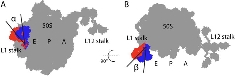
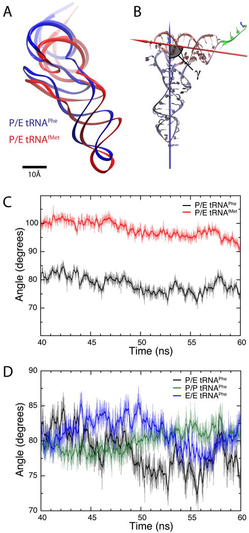
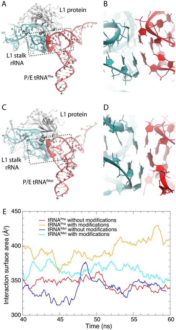
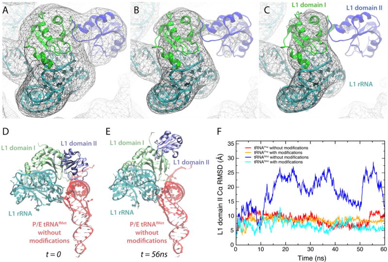
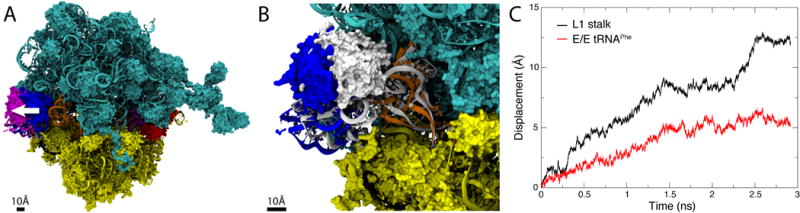
The ribosomal L1 stalk is a mobile structure implicated in directing tRNA movement during translocation through the ribosome. This article investigates three aspects of L1 stalk-tRNA interaction. First, by combining data from cryo electron microscopy, X-ray crystallography, and molecular dynamics simulations through the molecular dynamics flexible fitting method, we obtained atomic models of different tRNAs occupying the hybrid P/E state interacting with the L1 stalk. These models confirm the assignment of fluorescence resonance energy transfer states from previous single-molecule investigations of L1 stalk dynamics. Second, the models reconcile how initiator tRNA(fMet) interacts less strongly with the L1 stalk compared to elongator tRNA(Phe), as seen in previous single-molecule experiments. Third, results from a simulation of the entire ribosome in which the L1 stalk is moved from a half-closed conformation to its open conformation are found to support the hypothesis that L1 stalk opening is involved in tRNA release from the ribosome.


Figures

Fig. 1
Fig. 1
Schematic illustrating two rotation angles (α and β) that account for most of the observed motion of the L1 stalk as seen in cryo-EM-derived atomic models (see Table 4). Two conformations of the L1 stalk are shown (blue = closed; red = open). See Materials and Methods for details on how α and β were calculated.
Fig. 2
Fig. 2
RNA modifications highlighted on the (A) tRNAPhe and (B) tRNAfMet structures used in this study. The conformations shown correspond to the last frame of 60-ns MD simulation trajectories of tRNAs in the hybrid P/E state. On the right, secondary structure diagrams for each tRNA are given. Symbols for RNA modifications: s4U = 4-thiouridine; D = dihydrouridine; Ψ = pseudouridine; ms2i6A = 2-methylthio-N6-isopentenyladenosine; m7G = 7-methylguanosine; acp3U = 3-(3-amino-3-carboxypropyl)uridine; T = thymine; Cm = 2′-O-methylcytidine.
Fig. 3
Fig. 3
Simulated system. (A) Atomic model of the ribosome containing a hybrid P/E tRNA, obtained by flexibly fitting atomic structures into a cryo-EM reconstruction using MDFF (see Materials and Methods). (B) Simulated subsystem representing a P/E tRNA interacting with the L1 stalk. The simulated system is shown in color with the rest of the ribosome structure shown in light gray for reference. 50S = cyan; 30S = yellow; P/E tRNA = red; mRNA = black.
Fig. 4
Fig. 4
Structure of tRNAs inside the ribosome. (A) Relative position of P/E tRNAPhe (blue) and P/E tRNAfMet (red), calculated as an average from the last 20 ns of each MD simulation (all modified ribonucleosides included). (B) Definition of the angle γ formed between the two arms of the tRNA. One arm consists of the anticodon-stem loop, D loop and variable loop (ice blue), while the other is comprised of the acceptor stem and T loop (pink). The arrows show the principal axes of inertia corresponding to the smallest moments of inertia for each of the two arms. The residues in the single strand at the 3′ terminus (green) were not taken into account in the analysis. (C) Angle γ calculated for the last 20 ns for the simulation of P/E tRNAPhe (black) and P/E tRNAfMet (red). The thick lines show running averages with a 100-ps window. (D) Angle γ calculated for the last 20 ns for the simulations containing tRNAPhe in classical P/P (green) and E/E (blue) as well as hybrid P/E (black) states. The thick lines show corresponding running averages with a 100-ps window.
Fig. 5
Fig. 5
Per-residue root mean square fluctuations (RMSFs) for the individual tRNAs obtained from the last 20 ns of each trajectory. Panels A and B compare non-modified (black) with fully modified (red) P/E tRNAPhe and P/E tRNAfMet. Panel C compares modified P/E tRNAPhe (black) and P/E tRNAfMet (red). The blue triangle indicates the insertion within tRNAfMet with respect to tRNAPhe. Panel D shows a comparison of the RMSFs for the fully modified tRNAPhe in the P/E (black), P/P (red), and E/E (blue) states.
Fig. 6
Fig. 6
Stacking between P/E tRNA and L1 stalk bases. The figure shows structures obtained after 60 ns of MD simulations of (A, B) tRNAPhe and (C, D) tRNAfMet at the hybrid P/E site interacting with the L1 stalk. All RNA modifications were adopted in the simulations. Highlighted in surface representations are RNA residues involved in stacking interactions between the L1 stalk (G2112–A2169) and tRNA (G19–C56). The dashed boxes represent structural elements formed by sets of residues from both the L1 stalk (helix 77) and the tRNA's elbow involved in a series of stacking interactions. (E) Interaction surface area for residues involved in stacking interactions between tRNA and L1 stalk. Data calculated from the last 20 ns of each trajectory considering every 2 ps and applying a running average with a window size of 20 ps.
Fig. 7
Fig. 7
Interaction between U1851 (23S rRNA) and G70 (tRNA), observed in the fully modified P/E tRNAPhe simulation. (A) Part of helix 68 is shown, with a dashed circle highlighting the interaction with P/E tRNAPhe. (B) C3-G70-U1851 base triple is represented in detail. (C) Distance corresponding to the hydrogen bond between U1851 and G70, showing that this interaction forms after about 40 ns in the simulation containing fully modified P/E tRNAPhe and is stable thereafter.
Fig. 8
Fig. 8
Interdomain motion of ribosomal protein L1. (A, B, C) Cryo-EM density map (EMD 1363, Ref.7) contoured at increasing threshold levels, together with atomic model obtained via flexible fitting (see Materials and Methods). The images clearly show that L1's domain II (residues 69–159) has lower density than domain I, indicating interdomain motion. (D, E) In the simulation of P/E tRNAfMet without modified ribonucleosides, a significant movement of L1's domain II with respect to the rest of the L1 stalk is observed. Shown are snapshots from the MD trajectory that illustrate the observed range of motion. (A movie of the entire trajectory is available in Supplementary Data). (F) Cα RMSD of L1's domain II throughout each MD P/E tRNA trajectory after aligning the entire L1 protein with respect to domain I.
Fig. 9
Fig. 9
L1 stalk opening simulation. (A) Ribosome model corresponding to the state at which the E/E tRNA is released from the ribosome. The initial half-closed L1 stalk (blue) was moved to the open conformation (magenta) using MDFF in order to observe the effect of L1 stalk opening on the E/E tRNA. The white arrow illustrates L1 stalk opening. (B) Inset showing the initial structure of E/E tRNA and L1 stalk in white and the final structure in the same colors as in panel A. (C) Displacement of the center of mass of the L1 stalk and E/E tRNA elbow during the L1 stalk opening simulation.

Similar articles

Cited by

References

    1. Moazed D, Noller H. Intermediate states in the movement of transfer RNA in the ribosome. Nature. 1989;342:142–148. - PubMed
    1. Blanchard SC, Kim HD, Gonzalez RL, Puglisi JD, Chu S. tRNA dynamics on the ribosome during translation. Proc Natl Acad Sci USA. 2004;101:12893–12898. - PMC - PubMed
    1. Cornish PV, Ermolenko DN, Noller HF, Ha T. Spontaneous inter-subunit rotation in single ribosomes. Mol Cell. 2008;30:578–588. - PMC - PubMed
    1. Agirrezabala X, Lei J, Brunelle JL, Ortiz-Meoz RF, Green R, Frank J. Visualization of the hybrid state of tRNA binding promoted by spontaneous ratcheting of the ribosome. Mol Cell. 2008;32:190–197. - PMC - PubMed
    1. Julián P, Konevega AL, Scheres SHW, Lázaro M, Gil D, Wintermeyer W, Rodnina M, Valle M. Structure of ratcheted ribosomes with trnas in hybrid states. Proc Natl Acad Sci USA. 2008;105:16924–16927. - PMC - PubMed

Publication types

LinkOut - more resources