Review
. 2010:90:73-158.
doi: 10.1016/S0070-2153(10)90003-3.

Lung organogenesis

Affiliations
Review

Lung organogenesis

David Warburton et al. Curr Top Dev Biol. 2010.

Abstract

Developmental lung biology is a field that has the potential for significant human impact: lung disease at the extremes of age continues to cause major morbidity and mortality worldwide. Understanding how the lung develops holds the promise that investigators can use this knowledge to aid lung repair and regeneration. In the decade since the "molecular embryology" of the lung was first comprehensively reviewed, new challenges have emerged-and it is on these that we focus the current review. Firstly, there is a critical need to understand the progenitor cell biology of the lung in order to exploit the potential of stem cells for the treatment of lung disease. Secondly, the current familiar descriptions of lung morphogenesis governed by growth and transcription factors need to be elaborated upon with the reinclusion and reconsideration of other factors, such as mechanics, in lung growth. Thirdly, efforts to parse the finer detail of lung bud signaling may need to be combined with broader consideration of overarching mechanisms that may be therapeutically easier to target: in this arena, we advance the proposal that looking at the lung in general (and branching in particular) in terms of clocks may yield unexpected benefits.


Figures

Figure 3.1
Figure 3.1
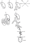
(A) The primitive lung anlage emerges as the laryngotracheal groove from the ventral surface of the primitive foregut at 5 weeks’ gestation in the human. (B) The primitive trachea separates dorsoventrally from the primitive esophagus as the two primary bronchial branches arise from the lateral aspects of the laryngotracheal groove at 5 or 6 weeks’ gestation in the human. (C) The embryonic larynx and trachea with the two primary bronchial branches are separated dorsoventrally from the embryonic esophagus at 6 weeks in the human. (D) The primitive lobar bronchi branching from the primary bronchi at 7 weeks in the human. (E) A schematic rendering of the airway at term in the human. The stereotypical first 16 airway generations are complete by 16 weeks in humans; between 16 and 24 weeks, further branching is nonstereotyped. Alveolarization begins about 20 weeks in humans and is complete by 7 years of age at the earliest. (After West, Burri, Warburton, and others).
Figure 3.2
Figure 3.2
Histology of mouse lung at characteristic stages of development. Embryonic mouse lung develops from pseudoglandular stage (E14.5) to canalicular stage (E16.5) and further terminal sac stage (E18.5 and P1). Neonatal mouse lungs undergo alveolarization, resulting in the formation of many septa (P14). Finally, a mature honeycomb-like structure with alveoli surrounding alveolar ducts conferring normal respiratory structure and function is formed, as observed in the adult. Scale bar: 100 μm.
Figure 3.3
Figure 3.3
How the airways can form in a sequential manner by reiterating a few, relatively simple sets of genetic instructions. In the upper panel, which is drawn after Warburton (2008), a master branch generator, a periodicity clock, and a bifurcator program are shown as controlling the layout of the mainstem and lobar branches. At embryonic day (E) 10.5, (A) the primary bronchial branch (1) forms, followed by (B) the development of the left upper-lobe branch (2) by E11, and then (C) the first two segmental branches of the left upper-lobe branch (2.2 and 2.3) form and the subsequent formation of branches 3–6 occurs by E12. The master branch generator is active throughout these events, and the inferred sites of action of the periodicity clock and bifurcator subroutines are shown. Then, in the lower panel, following the views of Metzger et al., (2008), a series of inferred genetic subroutines are shown, all driven by one master branch generator, shown as giving rise to domain or “bottle brush” branching along the lateral proximodistal axis of the main stem bronchi, which can then be rotated at right angles to give rise to a second rank of branches. Then, in subsequent rounds of branching, arising from the tips of the primary and secondary branches, it is shown how the same relatively simple periodicity generator, domain specifier, bifurcator, and rotator subroutines can give rise to apparently more complex patterns of peripheral branching to achieve an ever larger number of space filling terminal branches.
Figure 3.4
Figure 3.4
Schematic illustration of the variety of biochemical and biomechanical regulators of lung growth. The epithelium of the end-bud (yellow) encloses a fluid filled lumen (blue-green) in which oscillatory fluid flows are depicted (solid bidirectional arrows) that result from periodic peristaltic contractions (bi-directional curvilinear arrows) of airway smooth muscle (ASM: sample shown in brown running parallel and above the main epithelial trunk). The ASM derives from the FGF10+ precursor pool (seen in purple on the right), and these ASM progenitors are seen becoming more proximal (double-lined “=” at the top of the epithelial outline) relative to the growing epithelium. Examples of key biochemical signaling are given: FGF9 from the mesothelium (far right) regulates the FGF10+ mesenchyme (purple), which in turn interacts locally with epithelial Sprouty (SPRY2), BMP4, and Sonic Hedgehog (SHH) signaling (the latter two epithelial signals are shown in green and, due to space constraint, adjacent to the schematized epithelium). Epithelial Wnt signaling regulates fibronectin (FN) elaboration (shown as “xx”) in the cleft between epithelial branches. Epithelial VEGF signals to developing vasculature shown at the base of the figure. These vessels attract vascular smooth muscle precursors from the mesothelium (shown as “=” at the base of the figure). (See Color Insert.)
Figure 3.5
Figure 3.5
Subtypes of esophageal atresia (EA) and/or tracheo(T)–esophageal (E) fistula (TEF) with percentage frequency amongst EA-TEF cases. Type A: ‘Pure’ EA without TEF. Lower esophagus shown (LE); Type B: EA with TEF from proximal esophageal pouch but without any distal TEF; Type C: EA with distal TEF only (the most common variant); Type D: EA with both proximal TEF and distal TEF; Type E: TEF in the absence of EA. Laryngotracheal clefts (not shown here) are still rarer anomalies in which trachea and esophagus form a single lumen for a variable length. In severe variants, a combined tracheo-esophagus connects to the stomach whilst also giving rise to the main bronchi.
Figure 3.6
Figure 3.6
Excessive mesenchymal FGF signaling leads to overgrowth of tracheal rings. Wild-type and mutant tracheas are stained with Alcian blue. (A) Wild-type trachea at P0 exhibiting regular cartilage rings separated by noncartilaginous mesenchyme; (B) Fgfr2c+/Fgfr2b trachea at P0 showing excessive growth of the cartilage with absence of noncartilaginous mesenchyme; (C, D) high magnification of A and B, respectively. (See Color Insert.)
Figure 3.7
Figure 3.7
Vascular endothelial development in E12.5 mouse lung is shown in whole mount as a blue signal resulting from transgenic expression of Flk1-β-galactosidase (Flk-1nLacZ/+): pulmonary artery (PA); pulmonary vein (PV); cranial lobe (Cr); medial lobe (Med); caudal lobe (Ca); accessory lobe (Acc). (See Color Insert.)
Figure 3.8
Figure 3.8
Peristalsis occludes airway proximally whilst distending it distally. Sequential photomicrographs of cultured embryonic lung: in both panels the arrows outline proximal airway (open in the top picture but occluded by contraction in the lower one). The peristaltic airway occlusion’s distal effect is shown in a terminal bud, which rapidly rhythmically increases in size (the same ovoid outline is applied to both pictures). This distension and relaxation recurs with each wave.
Figure 3.9
Figure 3.9
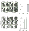
CaSR-evoked inhibition of branching and its dependence on Pulmonary lymphangitiic carcinomatosis (PLC) and phosphoinositide 3 (PI3) kinase signaling. Effect on branching of 1.05 mM (A, upper panels) or 1.7 mM (A, lower panels) Ca2+o in the absence (0.1% DMSO vehicle control; A left panels) or presence (A, right panels) of 5 μM of the PLC inhibitor, U73122. Quantification of branching at 48 h in the four conditions is shown in (B). Inhibition of PLC rescues suppression of branching evoked by 1.7 mM Ca2+o. Bars = 700 μm. Effect on branching of 1.05 mM Ca2+o (C, upper panels), 1.05 mM Ca2+o plus 30 nM R-568 (C, middle panels), and 1.7 mM Ca2+o (C, lower panels) in the absence (0.05% DMSO vehicle control) or presence of 12.5 μM of the PI3K inhibitor, LY294002. Bars = 750 μm. Quantification of branching at 48 h in the six conditions is shown in (D). The calcimimetic R-568 mimics the suppressive effect of high Ca2+o on branching, further implicating CaSR in the process. Note PI3 kinase inhibition rescues suppression of branching, whether evoked by high Ca2+o or calcimimetic. Adapted from Finney et al. (2008).
Figure 3.10
Figure 3.10
Smoking and genetics synergize to degrade lung function with age (modified after Fletcher and Peto, 1977; Shi, W. and Warburton, D. 2010). Wild-type lungs “grown in room air” achieve greatest capacity and remain healthy despite age-related degradation. Smoking exacerbates degradation of lung function even in the healthy. Genetic alterations decrease the potential to develop maximal lung capacity compared to wild type and smoking exacerbates lung degradation still further. A 0.5% loss of lung function per year is assumed for normal lungs and double that for smokers. Lungs with a genetic defect were assumed to have 95% of the growth rate of a normal lung. Growth rate is considered as a process of doublings, but the rate of doublings declines from maturity exponentially with age. This is expressed in a simple differential equation for modeling lung function; L(t):rateofchangeoflungfunction=rateofincreaseoflungfunctionrateofdeclineoflungfunction;dL/dt=rexp(matt)LaL, where mat is the maturation coefficient, r is the doubling coefficient, and a is the loss-of-function coefficient.

Similar articles

Cited by

References

    1. Acosta JM, Thebaud B, Castillo C, Mailleux A, Tefft D, Wuenschell C, Anderson KD, Bourbon J, Thiery JP, Bellusci S, Warburton D. Novel mechanisms in murine nitrofen-induced pulmonary hypoplasia: FGF-10 rescue in culture. Am J Physiol Lung Cell Mol Physiol. 2001;281:L250–257. - PubMed
    1. Adamson TM, Boyd RD, Platt HS, Strang LB. Composition of alveolar liquid in the foetal lamb. J Physiol. 1969;204:159–168. - PMC - PubMed
    1. Akiyama H, Lyons JP, Mori-Akiyama Y, Yang X, Zhang R, Zhang Z, Deng JM, Taketo MM, Nakamura T, Behringer RR, McCrea PD, de Crombrugghe B. Interactions between sox9 and beta-catenin control chondrocyte differentiation. Genes Dev. 2004;18:1072–1087. - PMC - PubMed
    1. Alcorn D, Adamson TM, Lambert TF, Maloney JE, Ritchie BC, Robinson PM. Morphological effects of chronic tracheal ligation and drainage in the fetal lamb lung. J Anat. 1977;123:649–660. - PMC - PubMed
    1. Ambros V. microRNAs: tiny regulators with great potential. Cell. 2001;107:823–826. - PubMed

Publication types

MeSH terms

Substances

LinkOut - more resources