Review
. 2010:90:263-90.
doi: 10.1016/S0070-2153(10)90007-0.

Building pathways for ovary organogenesis in the mouse embryo

Affiliations
Review

Building pathways for ovary organogenesis in the mouse embryo

Chia-Feng Liu et al. Curr Top Dev Biol. 2010.

Abstract

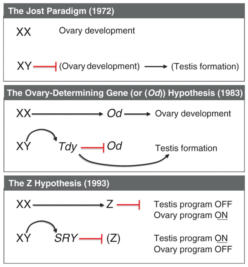
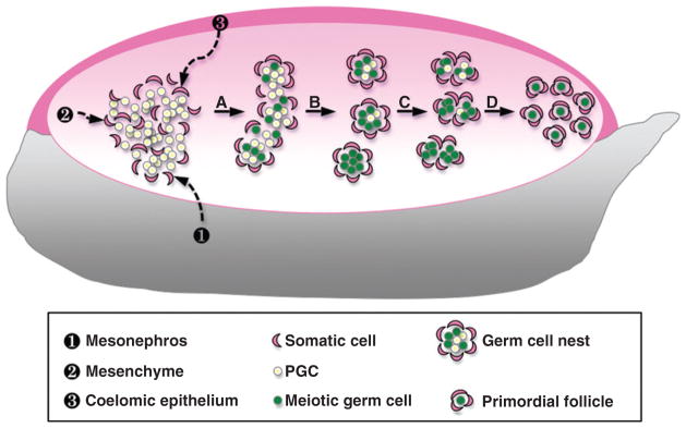
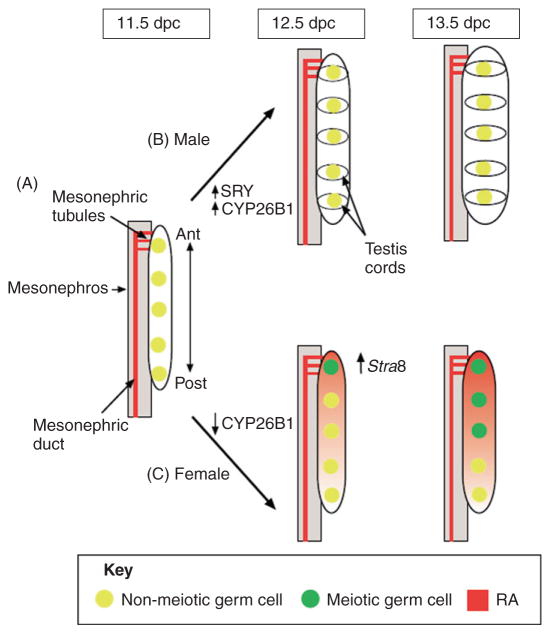
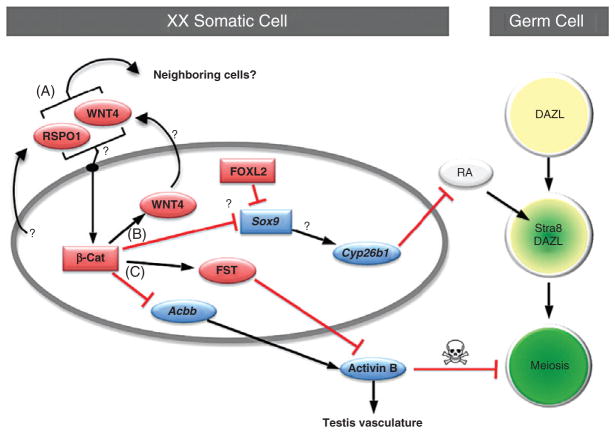
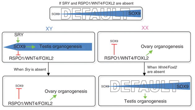
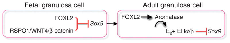
Despite its significant role in oocyte generation and hormone production in adulthood, the ovary, with regard to its formation, has received little attention compared to its male counterpart, the testis. With the exception of germ cells, which undergo a female-specific pattern of meiosis, morphological changes in the fetal ovary are subtle. Over the past 40 years, a number of hypotheses have been proposed for the organogenesis of the mammalian ovary. It was not until the turn of the millennium, thanks to the advancement of genetic and genomic approaches, that pathways for ovary organogenesis that consist of positive and negative regulators have started to emerge. Through the action of secreted factors (R-spondin1, WNT4, and follistatin) and transcription regulators (beta-catenin and FOXL2), the developmental fate of the somatic cells is directed toward ovarian, while testicular components are suppressed. In this chapter, we review the history of studying ovary organogenesis in mammals and present the most recent discoveries using the mouse as the model organism.


Figures

Figure 7.1
Figure 7.1
Evolution of the hypotheses for sex determination in eutherian mammals. In 1972, Jost extended his paradigm on sexually dimorphic development of the reproductive tracts to gonad differentiation and proposed that a putative “male organizer” prevents the gonadal primordium from developing into an ovary and forces it to become a testis. In 1983, Eicher and Washburn proposed that an ovary-determining gene (Od) initiates ovary differentiation in the XX individual. The testis-determining gene on the Y chromosome (or Tdy), which becomes active earlier than the Od gene, suppresses the Od gene in the XY individuals. The Z theory was proposed in 1993 by McElreavey et al. after the discovery of SRY gene. It was stated that the Z gene in the XX gonad inhibits the testis pathway, therefore leading to the progression of the ovary pathway. In the XY gonad, the SRY acts as an inhibitor of the Z gene, allowing the development of the testis.
Figure 7.2
Figure 7.2
Morphogenesis of the mouse fetal ovary. Once the PGCs migrate into and colonize the genital ridge, they coalesce with somatic cells which could be derived from three potential sources: ❶ the neighboring mesonephros, ❷ the existing mesenchyme, and/or ❸ the surrounding coelomic epithelium. (A) Somatic cells and PGCs form ovigerous cords. Female germ cells start to enter meiosis around 13.5 dpc. (B) Germ cell nests that are surrounded loosely by somatic cells begin to form. (C) Around the time of birth, somatic cells start to break down the germ cell nests by enclosing individual oocytes. (D) Breakdown of the germ cell cysts leads to the formation of the primordial follicles.
Figure 7.3
Figure 7.3
Regulation of germ cell entry into meiosis in the developing gonads. (A) At 11.5 dpc, PGCs are present in the genital ridge and RA is produced in the neighboring mesonephric duct and tubules. Cyp26b1 is expressed at low levels in the gonad of both sexes. The mesonephric tubules, which produce RA, are physically connected with the anterior (Ant) end of the gonad during this time. (B) In the male gonad, once Sry is expressed (~11.5 dpc), the RA-degrading enzyme Cyp26b1 expression is upregulated. The testis cords, which form around germ cell clusters around 12.5 dpc, might concentrate the enzyme in these regions, thereby protecting germ cells from the actions of RA. Germ cells in the male gonad therefore do not enter meiosis at 13.5 dpc. (C) In the female gonad, Cyp26b1 expression is detectable at 11.5 dpc, but disappears by 12.5 dpc. Germ cells at the anterior end of the gonad begin to express Stra8 at 12.5 dpc. By 13.5 dpc, female germ cells enter meiosis in an anterior-to-posterior (Post) wave. Germ cells at the anterior end of the gonad might be exposed to RA earlier than those at the posterior end, or the RA concentration might be greater at the anterior end than the posterior end [this figure is modified from Figure 2 in Bowles and Koopman (2007)].
Figure 7.4
Figure 7.4
Putative pathways for ovary organogenesis in the mouse embryo. (A) Two somatic cell-derived factors, R-spondin1 (RSPO1) and WNT4, activate synergistically or independently the canonical β-catenin (β-cat) pathway in XX somatic cells in an autocrine or paracrine manner. (B) β-catenin then induces expression of Wnt4 while suppresses expression of Sox9 (probably through the action of FOXL2) and its putative downstream target Cyp26b1. Without the presence of Cyp26b1, RA is not degraded and therefore induces Stra8 expression and meiosis in DAZL-positive germ cells. (C) β-catenin also induces expression of follistatin (Fst) and at the same time maintains a low expression of activin βB (Acbb). FST antagonizes the action of activin B, which is the protein product of Acbb. Lack of activin B ensures that no testis-specific vasculature forms and the survival of female germ cells is maintained. See text for more details. (See Color Insert.)
Figure 7.5
Figure 7.5
The 2010 version of sex determination hypothesis based on mouse genetic models. We propose that if both SRY and RSPO1/WNT4/FOXL2 are absent, the default status of gonads is testis, as a result of gradual increase of Sox9 expression. When both SRY and RSPO1/WNT4/FOXL2 are present as in the XY individual, SRY in the testis jumpstarts Sox9 expression, which subsequently suppresses the pro-ovary functions of RSPO1/WNT4/FOXL2. On the other hand, in the absence of Sry as in the XX individual, RSPO1/WNT4/FOXL2 prevent the rise of Sox9 and its ability to induce testis differentiation, therefore allowing the gonads to follow the ovarian path. When Sry is nonfunctional or lost, RSPO1/WNT4/FOXL2 synergistically suppresses Sox9 expression and facilitates ovary organogenesis. If the action of RSPO1/WNT4/FOXL2 is silenced in the XX individual, the default testis pathway arises despite the absence of Sry.
Figure 7.6
Figure 7.6
Maintenance of granulosa cell identity in fetal and adult ovaries. In the fetal ovary, granulosa cell identity is maintained in an estrogen-independent manner via the RSPO1/WNT4/β-catenin pathway and FOXL2, which together repress the expression of Sox9. This mechanism becomes estrogen-dependent after birth and in the adult ovary FOXL2 stimulates estrogen-producing enzyme aromatase and through the action of estrogen (E2) and their receptors (ERα/β) suppresses Sox9 and maintain the identity of granulosa cells.

Similar articles

Cited by

References

    1. Albrecht KH, Eicher EM. Evidence that Sry is expressed in pre-Sertoli cells and Sertoli and granulosa cells have a common precursor. Dev Biol. 2001;240:92–107. - PubMed
    1. Anderson R, Copeland TK, Scholer H, Heasman J, Wylie C. The onset of germ cell migration in the mouse embryo. Mech Dev. 2000;91:61–68. - PubMed
    1. Baltus AE, Menke DB, Hu YC, Goodheart ML, Carpenter AE, de Rooij DG, Page DC. In germ cells of mouse embryonic ovaries, the decision to enter meiosis precedes premeiotic DNA replication. Nat Genet. 2006;38:1430–1434. - PubMed
    1. Barbaro M, Cicognani A, Balsamo A, Lofgren A, Baldazzi L, Wedell A, Oscarson M. Gene dosage imbalances in patients with 46, XY gonadal DSD detected by an in-house-designed synthetic probe set for multiplex ligation-dependent probe amplification analysis. Clin Genet. 2008;73:453–464. - PubMed
    1. Barsoum IB, Bingham NC, Parker KL, Jorgensen JS, Yao HHC. Activation of the hedgehog pathway in the mouse fetal ovary leads to ectopic appearance of fetal Leydig cells and female pseudohermaphroditism. Dev Biol. 2009;329:96–103. - PMC - PubMed

LinkOut - more resources