. 2010 Aug 6;142(3):456-67.
doi: 10.1016/j.cell.2010.06.035.

PNPASE regulates RNA import into mitochondria

Affiliations

PNPASE regulates RNA import into mitochondria

Geng Wang et al. Cell. .

Abstract

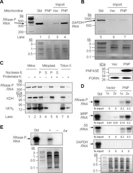
RNA import into mammalian mitochondria is considered essential for replication, transcription, and translation of the mitochondrial genome but the pathway(s) and factors that control this import are poorly understood. Previously, we localized polynucleotide phosphorylase (PNPASE), a 3' --> 5' exoribonuclease and poly-A polymerase, in the mitochondrial intermembrane space, a location lacking resident RNAs. Here, we show a new role for PNPASE in regulating the import of nuclear-encoded RNAs into the mitochondrial matrix. PNPASE reduction impaired mitochondrial RNA processing and polycistronic transcripts accumulated. Augmented import of RNase P, 5S rRNA, and MRP RNAs depended on PNPASE expression and PNPASE-imported RNA interactions were identified. PNPASE RNA processing and import activities were separable and a mitochondrial RNA targeting signal was isolated that enabled RNA import in a PNPASE-dependent manner. Combined, these data strongly support an unanticipated role for PNPASE in mediating the translocation of RNAs into mitochondria.


Figures

Figure 1
Figure 1. Deletion of Pnpt1 in Hepatocytes Impairs Mitochondrial Function
(A) Breeding strategy (top) and results (bottom) for attempting to generate a PNPASE KO mouse. (B) Hepatocyte-specific Pnpt1 KO (HepKO) in 4-week old mice. Top: QPCR for liver Pnpt1 expression using an exon 2 – exon 3 primer pair versus a primer pair within exon 28. Bottom: PNPASE immunoblot from 4-week old WT and HepKO mouse livers. (C) HepKO mitochondria have altered cristae. Left: TEM of 6-week old littermate livers shows circular, smooth HepKO IM cristae in contrast to linear, stacked cristae of WT mitochondria. Right: Analysis of cristae morphology in which a single normal cristae within a mitochondrion was scored as normal. Indet = indeterminate. (D) Decreased respiration in isolated HepKO mitochondria. Oxygen consumption (nmol/min/mg protein) for ETC complexes IV and II+III+IV was measured using an O2 electrode. Mitochondrial mass was determined by citrate synthase (CS) activity using a spectrophotometer. Respiratory activities are shown normalized to CS activity. (E) Decreased mature mtRNAs in HEK293 cells with RNAi to PNPT1. Transcripts were quantified relative to cytosolic GAPDH expression by QPCR from HEK293 cells 7d post-infection (nadir) with scramble (Scr) or PNPT1 RNAi retroviral constructs. See also Figure S1, S2, and S3.
Figure 2
Figure 2. HepKO Liver Mitochondria Do Not Efficiently Process mtRNA Precursors
(A) In organello protein synthesis. WT and HepKO mitochondria (100 μg) were treated with micrococcal nuclease S7, and in organello translation was performed using [35S]-MET. TOM40 immunoblot shows equivalent mitochondria in each assay. (B) RNA was isolated from WT and HepKO liver mitochondria followed by DNase I treatment to remove contaminating DNA. RT-PCR was performed for Cox1 and Cox2 with primers shown in the schematic (upper) and separated on a 1.5% agarose gel. (C) Northern blot of mtRNA from WT and HepKO mouse liver mitochondria using a Cox1 or Cox3 DNA probe. * marks larger precursor mtRNAs and the arrow shows the mature mtRNA. (D) Steady-state expression of nuclear- and mitochondrial-encoded proteins in WT and HepKO liver mitochondria. Equivalent nuclear-encoded protein expression shows that HepKO reduced mitochondria-encoded protein expression was not due to differing mitochondrial content between WT and HepKO liver cells. See also Figure S4.
Figure 3
Figure 3. RNase P RNA Binds to PNPASE and May Function in PNPASE-Dependent tRNA Processing
(A) Left- RNA was isolated from WT and HepKO liver mitochondria following nuclease treatment. RT-PCR was performed with primers that amplify nuclear-encoded RNase P RNA (212-bp). Right- QPCR analysis of RNase P RNA expression relative to TOM40 protein in isolated mitochondria. (B) PNPASE-HisPC (PNP) or TIM23-HisPC (TIM23) was purified from stably-transfected HEK293 cells. Candidate interacting RNAs that co-purified in the final eluate with PNPASE-HisPC and TIM23-HisPC were identified by primer-specific RT-PCR. T is the total lysate (0.3% of the reaction) before mitochondrial purification and B is the bound fraction. Note that only RNase P RNA bound to PNPASE-HisPC (lane 4). (C) Mitoplast extract (10 μg) was prepared from nuclease-treated, intact WT liver mitochondria. The extract was then treated with nuclease (+), as indicated, and then inactivated with EDTA and EGTA. The nuclease-treated or untreated extract was incubated with abutted tRNAs (tRNAHistRNASer) or a single tRNA (tRNALys) at 25°C for 10 or 30 min. RNA was separated on an urea-acrylamide gel and detected by autoradiography. A MORTALIN immunoblot shows equivalent mitoplast extract in each assay. (D) Mitoplast extract was prepared from nuclease-treated, intact WT or HepKO liver mitochondria. The enzymatic assay was performed as described for panel C. See also Figure S5.
Figure 4
Figure 4. PNPASE Augments RNase P, 5S rRNA, and MRP RNA Import into Yeast Mitochondria
(A) Upper- In vitro transcribed human RNase P RNA was incubated with yeast mitochondria expressing human PNPT1 (PNP) or an empty vector (Vec) control. Non-imported RNA was digested with nuclease and the imported RNA was detected by RT-PCR. PNPT1-expressing mitochondria without added RNase P RNA was included as a specificity control for import and RT-PCR (lane 2 – Std, 1% of the reaction). Lower- Control showing equivalent total mitochondrial nucleic acid in each reaction. (B) Upper- As in panel A, with cytosolic human GAPDH RNA used as a substrate. Middle- Control showing equivalent total mitochondrial nucleic acid in each reaction. Lower- Western blot showing PNPASE expression and PORIN immunoblot showing equivalent mitochondria in each import assay. (C) After import as in panel A, mitochondria were subjected to osmotic shock, fractionated by centrifugation into soluble (S) and pellet (P) fractions, followed by proteinase K and nuclease additions where indicated. The pellet fraction was solubilized with Triton X-100 to expose the matrix. Localization was determined by RT-PCR for RNase P RNA and immunoblot for KDH (matrix) and cyt b2 (IMS) proteins. (D) Upper- Radiolabeled RNase P, MRP, 5S rRNA, and GAPDH human RNAs were in vitro transcribed and then incubated with yeast mitochondria expressing PNPASE or an empty vector control. Non-imported RNA was digested with nuclease, followed by RNA isolation, separation on a urea acrylamide gel, and autoradiography. Import reactions were repeated with 1X and 2X amounts of RNA. Lower- Control showing equivalent total mitochondrial nucleic acid in each reaction. (E) Upper- As in panel A except that the mitochondrial membrane potential (Δψ) was dissipated prior to import. Lower- Control showing equivalent total mitochondrial nucleic acid in each reaction.
Figure 5
Figure 5. PNPASE Mutations that Inactivate RNA Processing do not Affect RNA Import or Stability
(A) Upper- Schematic for the positions of point mutations made in the PNPASE protein. Listed are the in vitro effects of mutations on 3’ polymerase and RNA degrading activities, from (Portnoy et al., 2008). Middle- Import reactions were performed as in Figure 4A. Radiolabeled RNase P RNA was incubated with isolated yeast mitochondria expressing an empty vector or the listed PNPASE constructs. Lower panels- Immunoblot of WT and point mutant PNPASE yeast transfectants used in panel A import assay. A PORIN immunoblot confirms the co-localization of PNPASE WT and mutants in yeast mitochondria. The assembly state of WT and point mutant PNPASE was determined by solubilization with 1% digitonin and separation on a 6-16% BN gel, followed by PNPASE immunoblot. (B) Upper- WT and S484A PNPASE IPs from yeast mitochondria were used to analyze RNA degradation properties. Lower- WT or S484A mutant PNPASE was incubated with radiolabeled RNase P RNA for 10 min at 25°C to assess degradation activity. The asterisk marks degradation products. (C) Left- Following in vitro import of radiolabeled RNase P RNA and nuclease treatment to remove non-imported RNA, mitochondria were incubated for up to 90 min at 25°C and aliquots removed at the indicated time points. The RNA was then resolved by urea-acrylamide gel electrophoresis. Right- RNase P RNA that was not degraded was quantified using FX imager; n = 3.
Figure 6
Figure 6. A Stem-Loop Structure Mediates PNPASE-Dependent RNA Import
(A) Schematic depiction of human RNase P RNA and deletion fragments. (B) Import of full length RNase P RNA into yeast mitochondria expressing PNPASE (PNP) or control (Vec) vectors, as in Figure 4A. (C) Import of the indicated RNase P RNA fragments. (D) Import of RNase P RNA fragments RPf3 and RPf4. (E) Import of human GAPDH mRNA or GAPDH mRNA with control (CR), MRP RNA, or RNase P RNA 20 nt sequences fused to the 5’ end, as shown in panel F. (F) The secondary structures and sequences of mitochondrial RNA targeting signals in RNase P (RP) and MRP (MRP) RNAs. A random sequence (CR) was used as a control. (G) Isolated mitochondria from HEK293 cells stably expressing IMS-localized PNPASE-HisPC or TIM23-HisPC (control) dual-tagged proteins were incubated with [32P]-CTP labeled CR-tRNAtrp or RP-tRNAtrp, followed by UV-cross linking, tag-IP, separation by SDS-PAGE, and autoradiography. See also Figure S6.
Figure 7
Figure 7. PNPASE Augments RNA Import into Yeast and Mammalian Mitochondria In Vivo
(A) Upper- Human RNase P RNA yeast expression construct is driven by the RPM1 RNA promoter, NME1. Lower-Mitochondria from yeast expressing human RNase P RNA and either PNPASE (PNP) or an empty vector (Vec) were isolated and treated with nuclease. RNA was then isolated from the total cell lysate or from nuclease-treated mitochondria (Mito) and analyzed by primer-specific RT-PCR. (B) QPCR for Cox1 and RNase P RNAs isolated from mitochondria in panel A, normalized to the total mitochondrial RNA obtained. (C) Radiolabeled, in vitro transcribed RNase P RNA was imported into mitochondria from MEF cell lines WT (expressing mouse PNPASE, mPNP), Pnpt1 knockout (KO), PNPT1 over-expression (expressing mPNP and hPNP), or Pnpt1 knockout plus PNPT1 over-expression (expressing hPNP). Upper panel is an immunoblot for mouse and human PNPASE expression. Middle panel is an immunoblot of β-ACTIN, a loading control. Lower panel is an autoradiogram of RNase P RNA import into isolated MEF mitochondria. (D) Radiolabeled, in vitro transcribed RNAs were incubated with WT or HepKO liver mitochondria for 10 min at 25°C. Non-imported RNA was removed with nuclease, followed by RNA isolation and separation on a urea- acrylamide gel. Import reactions were repeated with 1X and 2X amounts of synthesized RNAs. TOM40 immunoblot provides a mitochondrial loading control. See also Figure S7.

Comment in

References

    1. Alfonzo JD, Soll D. Mitochondrial tRNA import--the challenge to understand has just begun. Biol Chem. 2009;390:717–722. - PMC - PubMed
    1. Bonawitz ND, Clayton DA, Shadel GS. Initiation and beyond: multiple functions of the human mitochondrial transcription machinery. Mol Cell. 2006;24:813–825. - PubMed
    1. Carpousis AJ. The Escherichia coli RNA degradosome: structure, function and relationship in other ribonucleolytic multienzyme complexes. Biochem Soc Trans. 2002;30:150–155. - PubMed
    1. Chacinska A, Koehler CM, Milenkovic D, Lithgow T, Pfanner N. Importing mitochondrial proteins: machineries and mechanisms. Cell. 2009;138:628–644. - PMC - PubMed
    1. Chang DD, Clayton DA. A mammalian mitochondrial RNA processing activity contains nucleus-encoded RNA. Science. 1987;235:1178–1184. - PubMed

Publication types