. 2010 Aug 6;142(3):480-93.
doi: 10.1016/j.cell.2010.06.037.

Myc-nick: a cytoplasmic cleavage product of Myc that promotes alpha-tubulin acetylation and cell differentiation

Affiliations

Myc-nick: a cytoplasmic cleavage product of Myc that promotes alpha-tubulin acetylation and cell differentiation

Maralice Conacci-Sorrell et al. Cell. .

Abstract

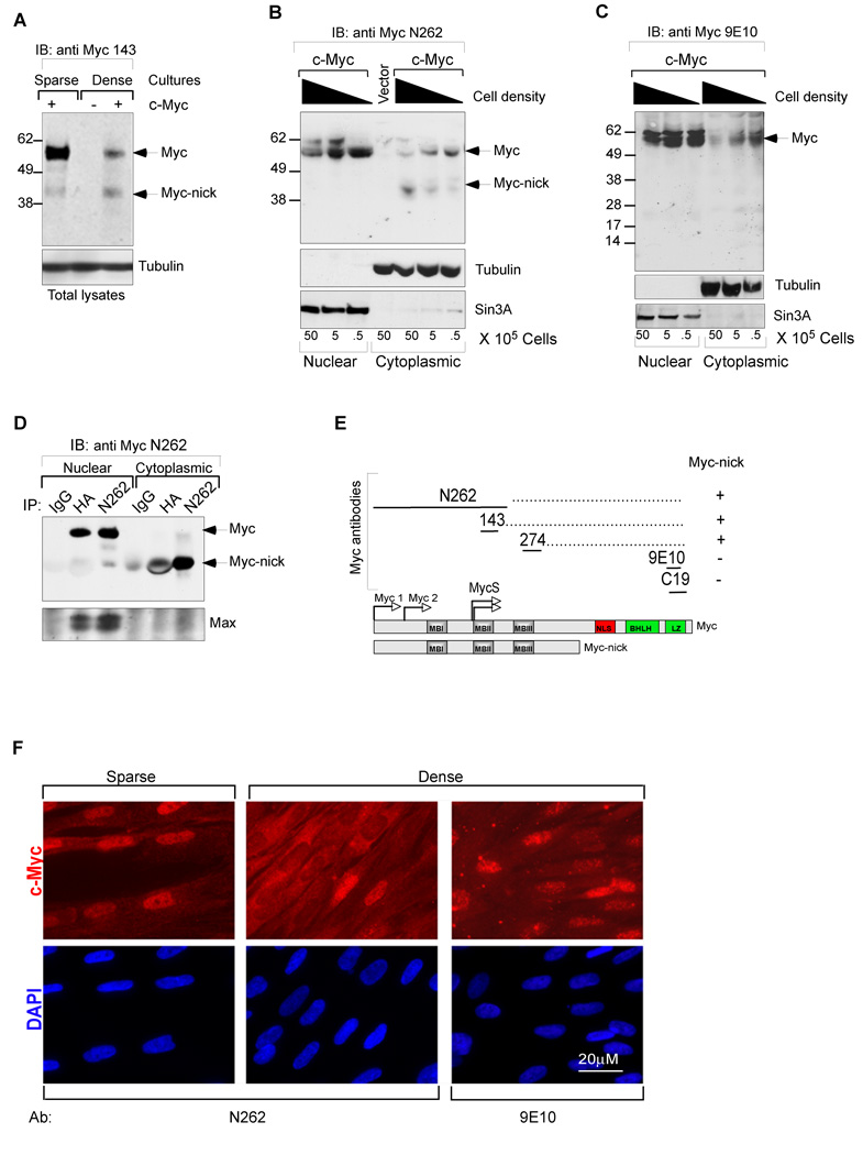
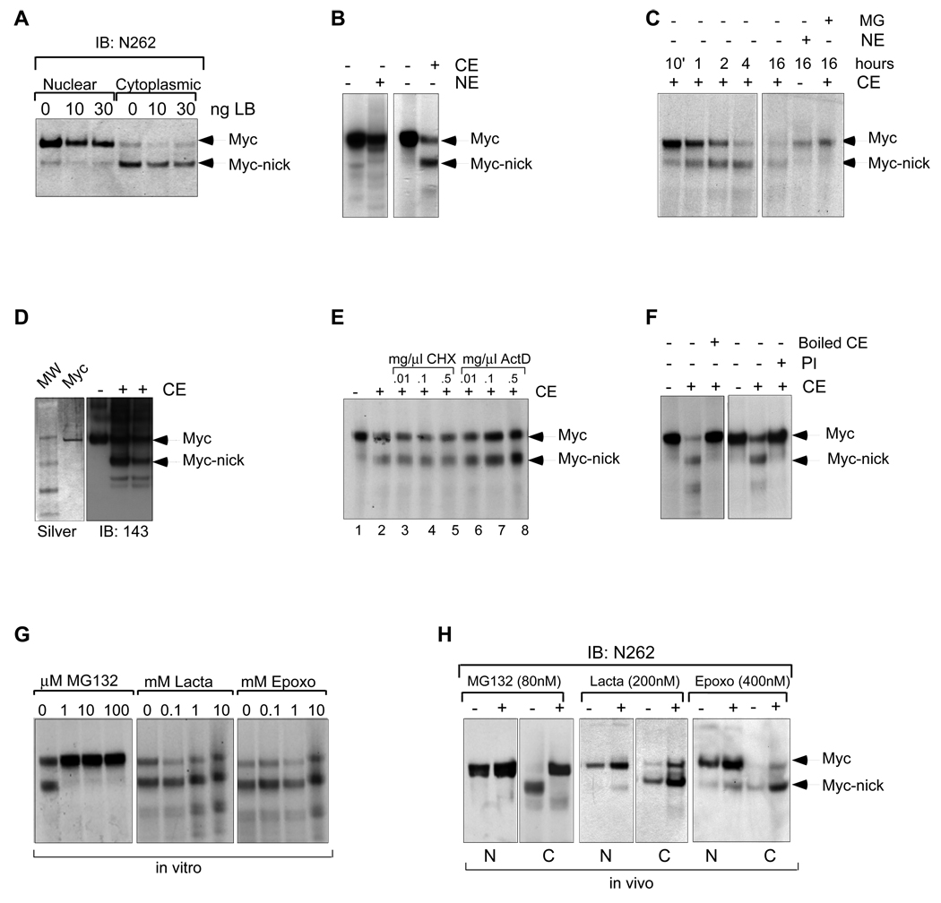
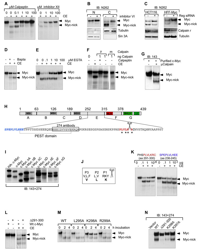
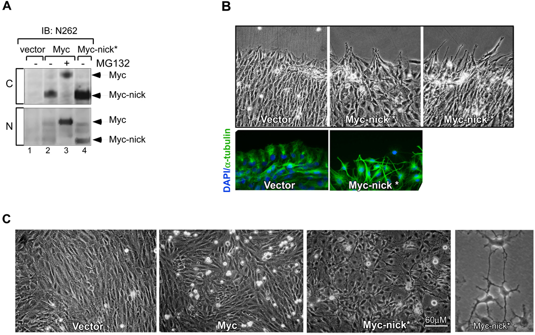
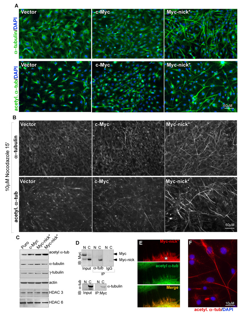
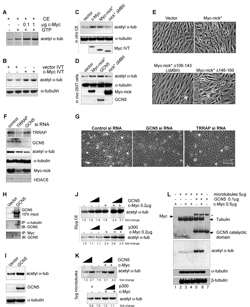
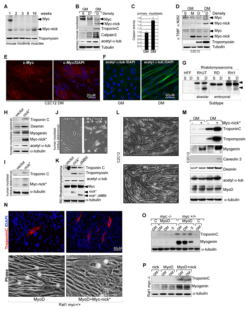
The Myc oncoprotein family comprises transcription factors that control multiple cellular functions and are widely involved in oncogenesis. Here we report the identification of Myc-nick, a cytoplasmic form of Myc generated by calpain-dependent proteolysis at lysine 298 of full-length Myc. Myc-nick retains conserved Myc box regions but lacks nuclear localization signals and the bHLHZ domain essential for heterodimerization with Max and DNA binding. Myc-nick induces alpha-tubulin acetylation and altered cell morphology by recruiting histone acetyltransferase GCN5 to microtubules. During muscle differentiation, while the levels of full-length Myc diminish, Myc-nick and acetylated alpha-tubulin levels are increased. Ectopic expression of Myc-nick accelerates myoblast fusion, triggers the expression of myogenic markers, and permits Myc-deficient fibroblasts to transdifferentiate in response to MyoD. We propose that the cleavage of Myc by calpain abrogates the transcriptional inhibition of differentiation by full-length Myc and generates Myc-nick, a driver of cytoplasmic reorganization and differentiation.


Figures

Figure 1
Figure 1. Identification of Myc-nick in the cytoplasm of cells grown at high density
(A) Total cell lysates of Rat1 myc-null fibroblasts infected with c-Myc, or empty retroviral vectors were prepared for western blot by adding boiling sample buffer. (B–C) Nuclear and cytoplasmic fractions of HFF cells expressing c-Myc were prepared 48h after plating at the indicated increasing densities. (D) Immunoprecipitation of HA-c-Myc (N-terminal tag) with anti-N262, anti-HA, and normal IgG from nuclear and cytoplasmic fractions of HFFs. Note that Max is only co-immunoprecipitated along with nuclear c-Myc. (E) Schematic representation of antibody mapping. (F) HFF cells infected with c-myc expressing retrovirus were cultured for 4 days after reaching confluency (middle and right panels) and compared with a subconfluent culture (left panel) by immunofluorescence using N262 and 9E10 antibodies. Nuclear (N) and cytoplasmic (C) fractions. See also Fig. S1.
Fig. 2
Fig. 2. Myc-nick is product of c-Myc cytoplasmic cleavage independent of the proteasome
(A) CRM1 dependant nuclear export is not involved in the formation or localization of Myc-nick. Rat1 myc-null cells infected with c-Myc were treated with leptomycin B (LB) for 4h before harvesting. (B) c-Myc gives rise to Myc-nick in vitro when incubated with cytoplasmic extracts. Radiolabeled IVT c-Myc was incubated for 2h with cytoplasmic (CE) or nuclear extracts (NE) from Rat1 c-myc null cells. (C) Time course of in vitro cleavage of c-Myc. (D) Recombinant c-Myc is cleaved in the presence of CE. 1 µg of recombinant c-Myc was incubated with 30µg of CE and processed for Western blot. (E) IVT c-Myc was incubated with CE for 4h in the presence of of Actinomycin D (ActD) or cyclohexamide (CHX). (F) The cleavage of c-Myc is inhibited by protease inhibitors and by heat inactivation. CE was boiled prior to incubation with IVT c-Myc and the Protease Inhibitor (PI) was added to the incubation mixture. (G,H) The cleavage of c-Myc is inhibited by MG132, but not by Lactacystein or Epoxomycin. (G) IVT c-Myc was incubated with CE, in the presence of increasing amounts of MG132, Lactacystein and Epoxomycin for 1h. (H) Rat1 myc-null cells expressing c-Myc were incubated with MG132, Lactacytein or Epoxomycin for 2h prior to harvesting. See also Fig. S2.
Fig. 3
Fig. 3. Myc-nick is generated by calpain cleavage of full-length Myc
(A) IVT c-Myc was incubated with CE for 1h in the presence of the calpain inhibitors calpeptin and calpain inhibitor XII. (B) Dense cultures of HFF cells infected with a c-Myc retroviral vector were incubated with Calpain inhibitor VI for 2h prior to harvesting. (C) siRNA for calpain regulatory subunit (Reg) reduces the formation of Myc-nick in HCT116 and HFF-Myc cells. (D) Rat1 myc-null cells were incubated for 2h in the presence of the calcium chelant Bapta, and then cytoplasmic extracts were prepared and incubated with IVT c-Myc (as in 2B). (E) IVT c-Myc was incubated with CE for 1h in the presence of increasing amounts of EGTA. (F) IVT c-Myc was incubated with purified recombinant μCalpain or mCalpain and r subunit for 1h on ice. (G) 100ng of purified μcalpain and r subunit was incubated with 2µg of recombinant c-Myc, for 30 min on ice. (H) Schematic representation of c-Myc protein indicating A–G deletion regions (deletion endpoints indicated above), putative calpain cleavage region in red. (I) c-Myc and the deletion mutants ΔA-ΔG were expressed in 293T cells and 48h later the presence of a Myc-nick-like protein in cytoplasmic extracts was determined. (J) Amino acid preference for calpain cleavage region according to Tompa, 2004. P1–P3 indicate the position of preferred residues in relation to the cleavage site, bold letters indicate the c-Myc calpain cleavage site. (K) IVT c-Myc was incubated with CE in the presence of a peptide that contains the potential calpain cleavage site (amino acids 291–300-in red) or a nearby sequence (amino acids 236–245-in blue). (L) WT and Δ291 –300 IVT c-Myc were incubated with CE for 1h (as in 2B). (M) IVT WT, L295A, K298A and K299A c-Myc were incubated with CE for the indicated time points. (N) Cleavage products produced from the indicated c-Myc mutants in 293T cells. See also Fig. S3.
Fig. 4
Fig. 4. The expression of Myc-nick* promotes changes in cell morphology
(A) Myc-nick* (1–298) is cytoplasmic and has the same apparent molecular weight as Myc-nick derived from full-length c-Myc (compare lanes 2 and 4, upper panel). Rat1 myc-null cells expressing empty vector, full-length c-Myc, and Myc-nick were fractionated into nuclear (N) and cytoplasmic (C) fractionations. (B) Myc-nick* expressing cells extend protrusions at the wound edge. A confluent monolayer of Rat1 myc-null cells expressing either vector or Myc-nick was scratched using a 100 µl tip and phase contrast images taken at 12h. (C) Rat1 myc-null cells expressing empty vector, c-Myc and Myc-nick at 14 days after selection. See also Fig. S4.
Fig. 5
Fig. 5. Myc-nick* cells display increased levels of acetylated (-tubulin and microtubule stabilization
(A) Immunofluorescence for α-tubulin (upper panels) and acetylated α-tubulin (lower panels) of Rat1 myc-null cells infected with empty vector, c-Myc or Myc-nick.* (B) As for (A) but incubated in the presence of nocodazole for 15 minutes prior to fixation. (C) Immunoblotting of Rat1 cell extracts using antibodies against the indicated proteins. (D) NE or CE of Rat1 myc-null cells expressing c-Myc were immunoprecipitated with anti α-tubulin or normal mouse IgG and immunoblotted for Myc (top panel), or immunoprecipitated with anti c-Myc N262 antibody, and immunoblotted with anti α-tubulin. (E) Immunofluorescence for Myc and acetylated α-tubulin in A431 lung epithelial cells cell transfected with Myc-nick (*). (F) Detail of Myc-nick expressing cell stained for acetylated α-tubulin. See also Fig. S5.
Fig. 6
Fig. 6. c-Myc and the HAT GCN5 promote α-tubulin acetylation
(A) CE was incubated with recombinant c-Myc and c-Myc dilution buffer; or (B) with c-Myc IVT or vector IVT for 1h at 37C. The samples were immunoblotted as indicated. (C) CE was incubated with IVT vector, c-Myc, Myc-nick and Myc-nick ΔMBII (Δ106–143) for 30’ at 37°C, then processed for for immunoblotting. (D) 293T cells were transfected with empty vector, GCN5, Myc-nick, Myc-nick ΔMBII and processed for immunoblotting after 48h. (E) Rat1 myc-null cells were infected as indicated and photographed 10 days after selection. (F–G) Rat1 myc-null cell expressing Myc-nick were transfected with 100nM of control, TRRAP or GCN5 siRNA and 76h later processed for immunoblotting (F) or photographed (G). (H) CE of 293T cells transfected with empty or GCN5 vectors were immunoprecipitated with anti-α-tubulin or anti-Myc (143+274) and immunoblotted for GCN5. (I) 293T cells were transfected with control or GCN5 expressing vectors and 48h later processed for immunoblotting. (J–L) GCN5 induces acetylation of α-tubulin. (J) CE were incubated with 200ng of Myc, 100ng or 300ng of GCN5 (upper panel) or 100ng or 500ng of p300 (lower panel). (K) Assembled microtubules were incubated with 100ng or 500ng of recombinant full-length GCN5 (upper panel) or p300 (lower panel) in the presence or absence of 200ng of purified c-Myc. (L) Purified assembled microtubules were incubated with recombinant c-Myc and GCN5-catalytic domain. The asterisk indicates a nonspecific bacterial band copurified with GCN5-catalytic domain. See also Fig. S6.
Fig. 7
Fig. 7. Myc-nick accelerates muscle cell differentiation
(A) Hindlimb muscles dissected from 1, 2, 3, 8, and 16 weeks old mice were processed for immunoblotting using N-terminal anti-Myc sera (anti 143+274). (B) Mouse primary myoblasts isolated from hindlimb muscles of 8-week old mice were cultured as sparse cultures for 24h or as dense cultures in the presence of either growth medium (GM) or differentiation medium (DM) for 3 days. Total cell extracts were immunoblotted for c-Myc (anti 143+274) and indicated proteins. (C) Mouse primary myoblasts were cultured in GM or DM for 3 days, lysed in buffer A, and total calpain activity was measured using Suc-LLVY-AMC synthetic substrate. (n=2. Calpain activity in DM was compared with calpain activity in GM (set to 1) +/− SEM). (D) C2C12 mouse myoblasts cultured at sparse (S), medium (M) or high densities (D). Dense cultures were harvested or switched to DM for 7 days. Total cell extracts were immunoblotted for c-Myc using antibodies against total c-Myc (N262) or against phosphorylated T58 Myc, a signal for Myc degradation. (E) C2C12 cells cultured in DM for 7 days and stained for endogenous c-Myc (anti-N262). (F) C2C12 cells grown in the presence of growth medium (GM) or differentiation medium (DM) for 5 days and stained with anti-acetylated α-tubulin. (G) Western blotting for Myc in Rhabdomyosarcoma cell lines grown as dense or sparse cultures for 3 days. (H−I) Human myoblasts expressing vector, or Myc-nick were cultured in DM and processed for immunoblotting after 4 days. (J) Human myoblasts expressing vector, or Myc-nick were cultured in DM and photographed after 2 days. (K) RD Rhabdomyosarcoma cells expressing vector, Myc-nick, or Myc-nick ΔMBII (Δ106–143) were grown in DM for 4 days and processed for immunobloting. (L) C2C12 cells expressing vector, or Myc-nick, were grown as dense cultures and stimulated to differentiate for 4 days and then photographed. (M) C2C12 cells expressing vector, or Myc-nick were grown to confluency and harvested or stimulated to differentiate for 3 days and then harvested. Total cell lysates were immunoblotted with antibodies against the indicated proteins. (N) Rat1 (myc+/+) cells expressing MyoD, or MyoD + Myc-nick were cultured in DM for 3 days and photographed (lower panels), or stained for Troponin C and DAPI (upper panels). (O) Rat1 (myc+/+) or Rat1 myc-null (myc−/−) cells expressing vector (lane C), or MyoD were cultured in GM, DM or serum free medium (lane -S) for 4 days and processed for immunobloting. (P) Rat myc null (myc−/−) cells expressing Myc-nick, MyoD, or MyoD + Myc-nick were grown in GM or DM for 3 days and processed for immunoblotting. See also Fig. S7.

References

    1. Abe K, Takeichi M. NMDA-receptor activation induces calpain-mediated beta-catenin cleavages for triggering gene expression. Neuron. 2007;53:387–397. - PubMed
    1. Alexandrova N, Niklinski J, Bliskovsky V, Otterson GA, Blake M, Kaye FJ, Zajac-Kaye M. The N-terminal domain of c-Myc associates with alpha-tubulin and microtubules in vivo and in vitro. Mol Cell Biol. 1995;15:5188–5195. - PMC - PubMed
    1. Arthur JS, Elce JS, Hegadorn C, Williams K, Greer PA. Disruption of the murine calpain small subunit gene, Capn4: calpain is essential for embryonic development but not for cell growth and division. Mol Cell Biol. 2000;20:4474–4481. - PMC - PubMed
    1. Bai MK, Costopoulos JS, Christoforidou BP, Papadimitriou CS. Immunohistochemical detection of the c-myc oncogene product in normal, hyperplastic and carcinomatous endometrium. Oncology. 1994;51:314–319. - PubMed
    1. Calcagno DQ, Guimaraes AC, Leal MF, Seabra AD, Khayat AS, Pontes TB, Assumpcao PP, De Arruda Cardoso Smith M, Burbano RR. MYC insertions in diffuse-type gastric adenocarcinoma. Anticancer Res. 2009;29:2479–2483. - PubMed

Publication types