. 2011 Jan;35(1):7-18.
doi: 10.1016/j.dci.2010.07.006. Epub 2010 Aug 11.

Molecular characterisation of Toll-like receptors in the black flying fox Pteropus alecto

Affiliations

Molecular characterisation of Toll-like receptors in the black flying fox Pteropus alecto

Christopher Cowled et al. Dev Comp Immunol. 2011 Jan.

Abstract

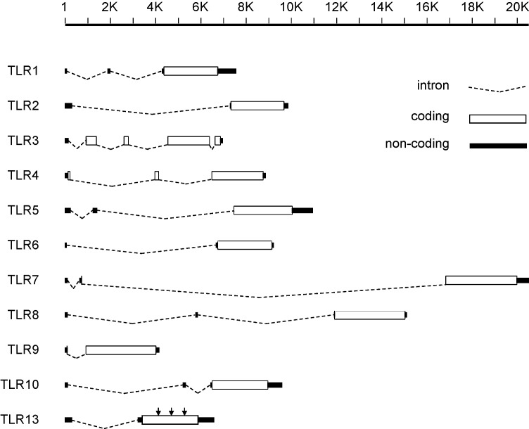
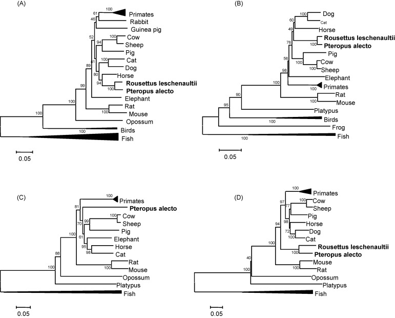
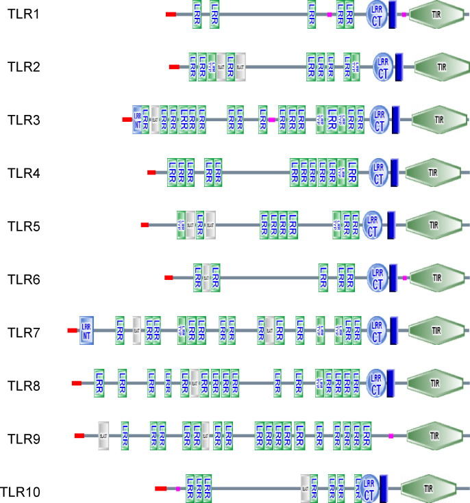
Bats are believed to be reservoir hosts for a number of emerging and re-emerging viruses, many of which are responsible for illness and mortality in humans, livestock and other animals. In other vertebrates, early responses to viral infection involve engagement of Toll-like receptors (TLRs), which induce changes in gene expression collectively leading to an "antiviral state". In this study we report the cloning and bioinformatic analysis of a complete set of TLRs from the black flying fox Pteropus alecto, and perform quantitative tissue expression analysis of the nucleic acid-sensing TLRs 3, 7, 8 and 9. Full-length mRNA transcripts from TLRs homologous to human TLRs 1-10 were sequenced, as well as a nearly intact TLR13 pseudogene that was spliced and polyadenylated. This prototype data can now be used to design functional studies of the bat innate immune system.


Figures

Fig. 1
Fig. 1
Predicted intron–exon organisation of pteropid bat TLRs 1–10 and TLR13. The intron–exon arrangements of pteropid bat TLRs were predicted by nucleotide alignment of P. alecto mRNA and P. vampyrus genomic DNA. Putative coding and non-coding sequences are drawn as light and dark rectangles respectively, while introns are indicated by dotted lines. P. alecto TLR13 is predicted to be a pseudogene due to the presence of three in-frame stop codons, as indicated by arrows. The figure is drawn to scale and distances are given in bp, however introns in TLRs 2, 4, 6, 7, 8 and 10 in the P. vampyrus genome assembly contained gaps so the actual sizes of these introns are estimates. The first exon of TLR5 was not represented in the P. vampyrus genome assembly and is therefore only assumed to be contiguous. Correspondingly, the first intron of TLR5 has been arbitrarily drawn at 1000 bp. In all other cases, intron–exon boundaries were clearly identified by homology in other species, and by the presence of splice site consensus sequences. In TLRs 7 and 9, the first coding exon contains only three coding nucleotides (ATG) that are hard to see in the diagram due to the scale.
Fig. 2
Fig. 2
Phylogenetic analysis of the virus-sensing TLRs 3, 7, 8 and 9 in P. alecto and other vertebrates. Phylogenetic trees generated from amino acid sequence alignments of (A) TLR3, (B) TLR7, (C) TLR8 and (D) TLR9 from P. alecto and other vertebrates. Refer to Table 2 for a list of accession numbers of protein sequences.
Fig. 3
Fig. 3
Predicted protein domain architecture of P. alecto TLRs 1–10. Protein domain architectures of P. alecto TLRs 1–10 were determined by SMART analysis. Signal peptides are shown in red, TM domains in dark blue and low complexity regions in pink. Other domains including LRR domains and TIR domains are labelled accordingly. (For interpretation of the references to colour in this figure legend, the reader is referred to the web version of the article.)
Fig. 4
Fig. 4
Transmembrane domain prediction in P. alecto TLRs 5 and 9. TM domain predictions were performed for (A) P. alecto TLR5 and (B) P. alecto TLR9. The output from two predictive algorithms (TMHMM, TMPRED) and three models of hydrophobicity (Kyte and Doolittle, Hopp and Woods, Surface exposure) are shown top to bottom for each protein. In the top plots (TMHMM), red peaks indicate probability of a TM domain, while the pink and blue lines indicate the best model (pink indicates outside the membrane while blue is inside the membrane). In the second plots, the dotted line is shown to indicate the predictive threshold–above the line is predicted to be a TM domain. The hydrophobicity plots are shown inverted–below the line represents hydrophobicity while above the line represents hydrophilicity. Putative TM domains are indicated by arrows. Outputs from the third predictive algorithm described in the text (HMMTOP) are not shown as they were text-based. *A second putative TM domain that was predicted in P. alecto TLR5 by all of the models tested. (C) Structural model of P. alecto TLR5 TIR domain based on human TLR2. The region in which a second putative TM domain was predicted by TMHMM and other methods is highlighted in red, and appears to form one of the central parallel β sheets. The C terminal helix of the TIR domain has been masked for clarity. (For interpretation of the references to colour in this figure legend, the reader is referred to the web version of the article.)
Fig. 5
Fig. 5
Structural modelling of P. alecto TLR ectodomains. Structural models of P. alecto TIR ectodomains (minus signal peptides) based on human TLR3: (A) TLR3, (B) TLR7, (C) TLR8, and (D) TLR9. The atypical N and C terminal LRRs identified by SMART are highlighted in red. The first and last LRR domains are numbered in each figure. All TLRs were slightly truncated at the ends by the modelling program. N and C terminals are indicated by capital letters, and arrows indicate regions that loop out from the main structures and may contain additional LRR domains. (For interpretation of the references to colour in this figure legend, the reader is referred to the web version of the article.)
Fig. 6
Fig. 6
Quantitative mRNA expression of TLRs 3, 7, 8 and 9 in P. alecto tissues. Tissue mRNA expression levels of: (A) TLR3, (B) TLR7, (C) TLR8, (D) TLR9. n = 3 individual apparently healthy wild-caught bats. Error bars represent standard deviation. Abbreviations: lymph node (L.N.), small intestine (Sm. int.), salivary gland (Sal. gland).

Similar articles

Cited by

References

    1. Altschul S.F., Gish W., Miller W., Myers E.W., Lipman D.J. Basic local alignment search tool. J. Mol. Biol. 1990;215:403–410. - PubMed
    1. Baker M.L., Tachedjian M., Wang L.F. Immunoglobulin heavy chain diversity in Pteropid bats: evidence for a diverse, highly specific antigen binding repertoire. Immunogenetics. 2010;62(3):173–184. - PMC - PubMed
    1. Barreiro L.B., Ben-Ali M., Quach H., Laval G., Patin E., Pickrell J.K., Bouchier C., Tichit M., Neyrolles O., Gicquel B., Kidd J.R., Kidd K.K., Alcais A., Ragimbeau J., Pellegrini S., Abel L., Casanova J.L., Quintana-Murci L. Evolutionary dynamics of human Toll-like receptors and their different contributions to host defense. PLoS Genet. 2009;5:e1000562. - PMC - PubMed
    1. Bieback K., Lien E., Klagge I.M., Avota E., Schneider-Schaulies J., Duprex W.P., Wagner H., Kirschning C.J., Ter Meulen V., Schneider-Schaulies S. Hemagglutinin protein of wild-type measles virus activates toll-like receptor 2 signaling. J. Virol. 2002;76:8729–8736. - PMC - PubMed
    1. Blehert D.S., Hicks A.C., Behr M., Meteyer C.U., Berlowski-Zier B.M., Buckles E.L., Coleman J.T., Darling S.R., Gargas A., Niver R., Okoniewski J.C., Rudd R.J., Stone W.B. Bat white-nose syndrome: an emerging fungal pathogen? Science. 2009;323:227. - PubMed

Publication types

Substances

Associated data