Review
. 2010 Sep;26(9):415-23.
doi: 10.1016/j.tig.2010.06.006. Epub 2010 Aug 6.

Psoriasis genetics: breaking the barrier

Affiliations
Review

Psoriasis genetics: breaking the barrier

Elisha D O Roberson et al. Trends Genet. 2010 Sep.

Abstract

Psoriasis is a common incurable inflammatory skin disease affecting 2-3% of the European population. Psoriatic skin contains large numbers of immune cells which produce many cytokines, chemokines and inflammatory molecules. The epidermis divides much faster than normal and has a defective outer layer or barrier which under normal circumstances protects from infection and dehydration. Psoriatic skin is characterized by a distinct set of inflammation and epidermal proliferation and differentiation markers, and it has been unclear whether the genetic basis of psoriasis reflects defects of the immune system or of the skin. One genetic determinant lies within the major histocompatibility complex class 1 region. Genome-wide association studies have revealed genetic susceptibility factors that play a role in the formation of immune cells found in psoriasis lesions. Others affect epidermal proliferation and skin barrier formation. Hence, genetic components of both the immune system and the epidermis can predispose to disease.


Figures

Figure 1
Figure 1
Different forms of psoriasis. (a) Plaque psoriasis is characterized by patches of inflamed skin with silvery scales. These lesions mostly target the elbows, knees and trunk. (b) Flexural (inverse) psoriasis is characterized by painful smooth, red and inflamed lesions in natural skin folds such as the inside of elbows and knees. (c) Seborrheic psoriasis targets areas such as the scalp or eyebrows with that have an oily appearance. (d) Guttate psoriasis lesions are punctate inflamed dots appearing in children or adolescents following infection with Streptococci bacteria. Guttate psoriasis in contrast to other forms is not chronic and typically resolves on its own. (e) Pustular psoriasis is more common in adults than in children. The lesions are numerous skin eruptions that are filled with sterile pus. (f) Non-pustular palmar-plantar psoriasis. (g) Nail psoriasis can lead to pitting and clouding of the nails, sometimes with total loss of the nail bed.
Figure 2
Figure 2
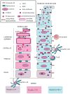
Diagram of normal and psoriatic involved epidermis found in lesions (not to scale). Normal epidermis contains approximately 10 cell layers made up of the basal layer, spinous layer, granular layer, and the cornified layer or stratum corneum (SC). The SC is constantly being sloughed off and replenished via proliferation in the basal layer. Keratin proteins including Keratins 5/14, 1/10 and 6/16/17 are generated in large amounts and represent the bulk of the keratinocyte weight [86]. S100 proteins such as S100A7 and S100A11 are present in the basal and spinous layers in normal epidermis. They appear in the nucleus and cytoplasm in basal cells but are associated with the plasma membrane in spinous cells in normal and psoriatic tissue[87]. S100A7 is also termed psoriasin and has antimicrobial functions [88]. Terminal differentiation proteins are made in the granular layer where nuclei break down, keratins condense, and cornified envelope (CE) proteins are cross-linked by transglutaminase. Mature keratinocytes in the cornified layer are known as corneocytes, lack nuclei and are flattened and condensed. The space between cells is filled neutral lipids that have been secreted from lamellar granules (LG) into the intercellular spaces of the upper granular layer to form intracellular lipid lamellae. The resultant organization of lipid and corneocytes protects the organism from infection and dehydration and has been compared to bricks and mortar [89]. Defensins are stored in lamellar granules and extruded with lipids into the intracellular space of normal skin [90]. At the same time desmosomes are transformed into corneodesmosomes by insertion of the protein corneodesmosin into the adhesive portion of these structures. In psoriasis lesions the granular layer is often absent, and corneocytes retain their nuclei (parakeratosis). The SC is thicker and disorganized. Components of the CE are also prematurely synthesized in the spinous layer. In psoriasis lesions neutral lipids are not secreted in a normal fashion into the extracellular space. This leads to a defective water/vapor barrier and the shedding of strateum corneum fragments in large sheets called scales or flakes in psoriasis plaques. In psoriasis antimicrobial peptides such as S100A7 and beta-defensin are highly upregulated. S100A7 is Connexin 26 (Cx26) is a gap junction protein that is also highly upregulated in psoriasis [48]. Transgenic over-expression of Cx26 in mouse epidermis keeps wounded epidermis in a hyper-proliferative state and blocks the transition to remodeling [81]. Its upregulation also leads to infiltration of immune cells.
Figure 3
Figure 3
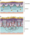
Comparison of immune cells in normal versus psoriatic skin illustrating how genetic risk factors could trigger and perpetuate epidermal inflammation and proliferation. (a) In normal skin there are a number of resident cells of the immune system. These include specialized dendritic cells (DCs) called Langerhan’s cells, as well as skin homing T cells and neutrophils. (b) In psoriasis lesions DCs and keratinocytes can act as antigen presenting cells (APCs) to lymphocytes via their major histocompatibility complex (MHC) proteins (see Box 1). Such an initiating trigger could lead to enhanced production of IL12 and IL23 by DCs in genetically susceptible individuals. In psoriasis lesions, a subset of DCs express high levels of tumour necrosis factor (TNF) and the enzyme inducible nitric oxide synthase (iNOS). These are termed TIP-DCs (TNF and iNOS-producing DCs). It is thought that these DCs produce the cytokines IL-23 and IL-20, which have the potential to activate T cells and keratinocytes, respectively [24]. IL23 triggers differentiation of Th17 cells following binding to the IL23R on naïve T cells. Production of Th17 cells would be enhanced by the presence of the IL23R genetic risk factor. TNF, gamma interferon, Th1 and Th17 cytokines induce keratinocytes in psoriasis lesions to synthesize numerous proteins that can attract the array of leukocytes found in lesional epidermis. This includes beta-defensins, and the S100A7 protein also known as psoriasin. Neutrophils accumulate in small aggregates in the cornified epidermis in the form of Munro abscesses. IL-22 cytokines act on keratinocytes to increase proliferation [46].

Similar articles

Cited by

References

    1. Bhalerao J, Bowcock A. The genetics of psoriasis: a complex disorder of the skin and immune system. Hum Mol Genet. 1998;7:1537–1545. - PubMed
    1. Boyman O, et al. Spontaneous Development of Psoriasis in a New Animal Model Shows an Essential Role for Resident T Cells and Tumor Necrosis Factor-α. The Journal of Experimental Medicine. 2004;199:731–736. - PMC - PubMed
    1. Arnett FC, et al. Psoriasis and psoriatic arthritis associated with human immunodeficiency virus infection. Rheumatic Diseases Clinics of North America. 1991;17:59–78. - PubMed
    1. Rongioletti F, et al. Psoriasis Induced or Aggravated by Drugs. The Journal of Rheumatology. 2009;83:59–61. - PubMed
    1. Nograles KE, et al. New insights into the pathogenesis and genetics of psoriatic arthritis. Nat Clin Pract Rheum. 2009;5:83–91. - PMC - PubMed

Publication types

Substances