. 2010 Oct 15;285(42):32038-46.
doi: 10.1074/jbc.M110.148452. Epub 2010 Aug 6.

Dependence on vitamin K-dependent protein S for eukaryotic cell secretion of the beta-chain of C4b-binding protein

Affiliations

Dependence on vitamin K-dependent protein S for eukaryotic cell secretion of the beta-chain of C4b-binding protein

Sofia Carlsson et al. J Biol Chem. .

Abstract

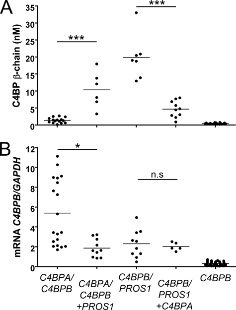
The anticoagulant vitamin K-dependent protein S (PS) circulates in plasma in two forms, 30% free and 70% being bound to the complement regulatory protein C4b-binding protein (C4BP). The major C4BP isoform consists of 7 α-chains and 1 β-chain (C4BPβ(+)), the chains being linked by disulfide bridges. PS binds to the β-chain with high affinity. In plasma, PS is in molar excess over C4BPβ(+) and due to the high affinity, all C4BPβ(+) molecules contain a bound PS. Taken together with the observation that PS-deficient patients have decreased levels of C4BPβ(+), this raises the question of whether PS is important for secretion of the β-chain from the cell. To test this hypothesis, HEK293 cells were stably and transiently transfected with β-chain cDNA in combinations with cDNAs for PS and/or the α-chain. The concentration of β-chains in the medium increased after co-transfection with PS cDNA, but not by α-chain cDNA, suggesting secretion of the β-chains from the cells to be dependent on concomitant synthesis of PS, but not of the α-chains. Thus, β-chains that were not disulfide-linked to the α-chains were secreted in complex with PS, either as monomers or dimers. Pulse-chase demonstrated that the complexes between PS and β-chain were formed intracellularly, in the endoplasmic reticulum. In conclusion, our results demonstrate that successful secretion of β-chains depends on intracellular complex formation with PS, but not on the α-chains. This provides an explanation for the decreased β-chain levels observed in PS-deficient patients.


Figures

FIGURE 1.
FIGURE 1.
Properties of liver cell lines HepG2, MIHA, and Hep3B. Protein concentrations (open bars) of C4BP α-chain, C4BP β-chain and PS, and mRNA levels (gray bars) of C4BPA, C4BPB and PROS1 were determined in HepG2 (A), MIHA (B), and Hep3B (C) cells. The protein concentrations were measured in cell medium collected from confluent cells. The mRNA content in the cells was detected by qRT-PCR and presented as the ratio between the gene of interest and the housekeeping gene (GAPDH). The bars correspond to the mean ± S.D. (error bars) value of at least three independent experiments.
FIGURE 2.
FIGURE 2.
Co-expression of PROS1 and C4BPB increases the expression of C4BP β-chain. HEK293 cells were stably transfected with the constructs indicated below each data set; C4BPA/C4BPB and C4BPB/PROS1 were in the vector pBudCE4.1, whereas C4BPA, C4BPB, and PROS1 were in pcDNA3. Multiple clones were picked and continuously cultured. A, concentrations of C4BP β-chain, PS (supplemental Fig. S2A), and C4BP α-chain (supplemental Fig. S2B) were measured in medium collected from confluent cells. B, mRNA levels of C4BPB, PROS1 (supplemental Fig. S3A), and C4BPA (supplemental Fig. S3B) were measured by qRT-PCR. The statistical significance of differences is indicated by horizontal lines; n.s., not significant. *, p < 0.05; ***, p < 0.001.
FIGURE 3.
FIGURE 3.
C4BP β-chain expression is stimulated by simultaneous synthesis of PS whereas miniPS and C4BP α-chain have no effect. HEK293 cells stably transfected with C4BPB in pcDNA3 were transiently transfected with PROS1, miniPS, cDNA, C4BPA, or empty pcDNA3 vector. Concentrations of C4BP β-chain (A), PS (B), and C4BP α-chain (supplemental Fig. S4) in the cell medium were measured 48 h after transfection. ***, p < 0.001.
FIGURE 4.
FIGURE 4.
C4BP β-chain expression stimulated by simultaneous synthesis of PS with intact C terminus. HEK293 cells stably transfected with C4BPA/C4BPB in pBudCE4.1 were transiently transfected with wild type PROS1, PS-CTERM, Gas6, a PS/Gas6 chimeric protein containing the Gas6 C terminus, or empty pcDNA3 vector. The concentrations of total C4BP and C4BP β-chain were measured in cell medium 48 h after transfection. The dots represent the fold change in β-chain concentration compared with pcDNA3, after normalization to total C4BP concentration in the same sample. Results that differed significantly from the pcDNA3 sample are marked with ** (p < 0.01) and *** (p < 0.001).
FIGURE 5.
FIGURE 5.
SDS-PAGE analysis of proteins from barium absorption. Samples (2 μg of protein/lane) were applied to a 4–15% gradient gel, electrophoretically separated, and silver stained. Proteins were eluted from the α-chain monoclonal column MK104 after application of barium citrate supernatant (S) or absorbed proteins (A) and can be compared with plasma-purified C4BP/PS (ctrl).
FIGURE 6.
FIGURE 6.
Western blot analysis of proteins eluted in stepwise purification. Eluted samples (100 ng of protein/lane) from the columns MK104 (binding C4BP α-chain), MK36 (binding C4BP β-chain), and MK54 (binding PS) or control (Ctrl) PS·C4BP purified from plasma, were applied to a 5% (B, left panel) or 10% SDS-PAGE, electrophoretically separated and transferred to a PVDF membrane. The proteins were detected with in-house mouse mAb against C4BP β-chain (A), in-house rabbit polyclonal antibody recognizing C4BP α-chains (B), and in-house mouse mAb against PS (unreduced sample) and a commercial rabbit polyclonal antibody (Dako) recognizing reduced PS (C). Adequate HRP-conjugated secondary antibodies were used to enable visualization with the ECL technique. Samples in the right panel were reduced by the addition of DTT.
FIGURE 7.
FIGURE 7.
Complex between C4BP β-chain and PS is formed inside the cells. HEK293 cells expressing PS, C4BP β-chain, and α-chain were metabolically labeled for 30 min (0 sample) and chased for 30 min, 90 min, or overnight (ON). At the depicted time points the cells were lysed, and the cell medium was harvested. Proteins in the cell lysates and medium were immunoprecipitated with a rabbit polyclonal antibody binding to either PS or C4BP, and proteins were separated with SDS-PAGE (10% or 5% separation gel) and transferred to a PVDF membrane. The bands on the membrane were visualized with a PhosphorImager FLA-3000.

Similar articles

Cited by

References

    1. Dahlbäck B., Villoutreix B. O. (2005) FEBS Lett. 579, 3310–3316 - PubMed
    1. Ndonwi M., Tuley E. A., Broze G. J., Jr. (2010) Blood 116, 1344–1351 - PMC - PubMed
    1. Hackeng T. M., Maurissen L. F., Castoldi E., Rosing J. (2009) J. Thromb. Haemost. 7, Suppl. 1, 165–168 - PubMed
    1. Dahlbäck B., Stenflo J. (1981) Proc. Natl. Acad. Sci. U.S.A. 78, 2512–2516 - PMC - PubMed
    1. Maurissen L. F., Thomassen M. C., Nicolaes G. A., Dahlbäck B., Tans G., Rosing J., Hackeng T. M. (2008) Blood 111, 3034–3041 - PubMed

Publication types

LinkOut - more resources