. 2010 Oct;9(10):1441-54.
doi: 10.1128/EC.00087-10. Epub 2010 Aug 6.

Eisosome organization in the filamentous ascomycete Aspergillus nidulans

Affiliations

Eisosome organization in the filamentous ascomycete Aspergillus nidulans

Ioannis Vangelatos et al. Eukaryot Cell. 2010 Oct.

Abstract

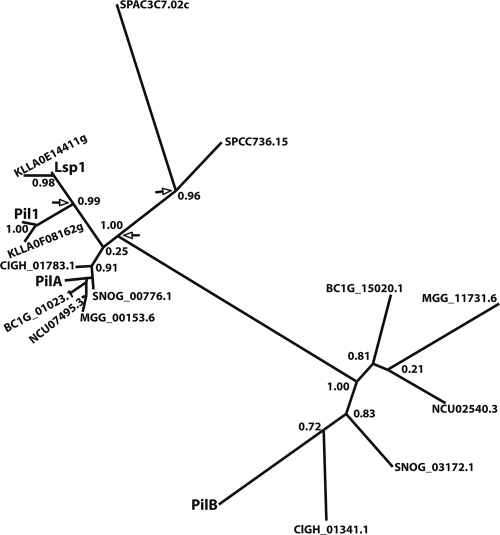
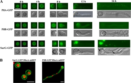
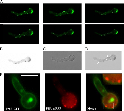
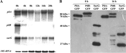
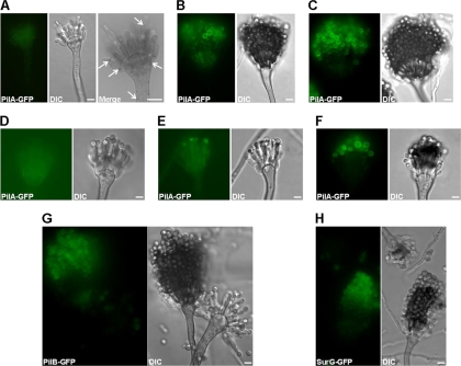
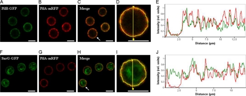
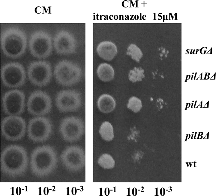
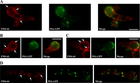
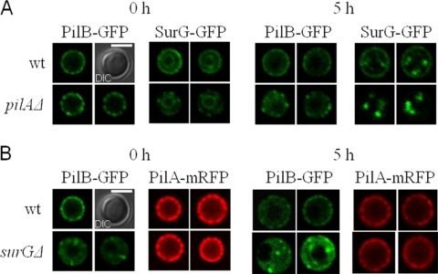
Eisosomes are subcortical organelles implicated in endocytosis and have hitherto been described only in Saccharomyces cerevisiae. They comprise two homologue proteins, Pil1 and Lsp1, which colocalize with the transmembrane protein Sur7. These proteins are universally conserved in the ascomycetes. We identify in Aspergillus nidulans (and in all members of the subphylum Pezizomycotina) two homologues of Pil1/Lsp1, PilA and PilB, originating from a duplication independent from that extant in the subphylum Saccharomycotina. In the aspergilli there are several Sur7-like proteins in each species, including one strict Sur7 orthologue (SurG in A. nidulans). In A. nidulans conidiospores, but not in hyphae, the three proteins colocalize at the cell cortex and form tightly packed punctate structures that appear different from the clearly distinct eisosome patches observed in S. cerevisiae. These structures are assembled late during the maturation of conidia. In mycelia, punctate structures are present, but they are composed only of PilA, while PilB is diffused in the cytoplasm and SurG is located in vacuoles and endosomes. Deletion of each of the genes does not lead to any obvious growth phenotype, except for moderate resistance to itraconazole. We could not find any obvious association between mycelial (PilA) eisosome-like structures and endocytosis. PilA and SurG are necessary for conidial eisosome organization in ways that differ from those for their S. cerevisiae homologues. These data illustrate that conservation of eisosomal proteins within the ascomycetes is accompanied by a striking functional divergence.


Figures

Fig. 1.
Fig. 1.
Eisosomal proteins in the subphylum Pezizomycotina. Phylogenetic unrooted tree, including the Pil1 (YGR086C) and Lsp1 (YPL004C) proteins of S. cerevisiae; the two putative eisosomal proteins of the second member of the Saccharomycotina, Kluvermyces lactis (KLLA); a member of the Taphrinomycotina, S. pombe (SPAC3C7.02c, SPCC736.15); and the PilA and PilB homologues (see text) of a number of representative fungi of the Pezizomycotina. The proteins of A. nidulans (Euriotiomycetes) referred to are PilA (ANID_05217.1) and PilB (ANID_03931.1) Other species referred to are Botritis cynerea (BC) (Leotiomycetes), Coccidioides immitis (CIGH; Euriotiomycetes), Magnaporthe grisae (MGG), Neurospora crassa (NCU; Sordariomycetes), and Stegonospora nodorum (SNOG; Dothideomycetes), followed by their systematic numbers in the relevant databases. Alignments were carried out with T-Coffee, curated with the Phylogeny site internal curation program (see Materials and Methods), and the tree was obtained with the maximum-likelihood program alLRT-PhyML (; http://www.phylogeny.fr/version2_cgi/alacarte.cgi) and redrawn after a Drawtree image. The digits at the nodes represent the results of the approximate likelihood ratio test and are nonparametric branch support values (http://atgc.lirmm.fr/alrt/). The PilA clade is clustered with all the homologues of the Saccharomycotina (approximate likelihood ratio test value = 1), but note the weak support (approximate likelihood ratio test value = 0.25) of the node where the PilA clade branches out from the homologues in the Saccharomycotina; in trees (data not shown) where many more species of Saccharomycotina and Pezizomycotina are included, the topology is maintained and the approximate likelihood ratio test value increases to >0. 80. For reasons of space, we do not show the branch support approximate likelihood ratio test values within the PilA clade; all nodes with the exception of the node where PilA branches out have strong support (approximate likelihood ratio test value > 0.80); the node where PilA branches out has an approximate likelihood ratio test value of 0.43. While we show only representative species in this tree, the topology of a tight cluster of PilA homologues and a looser cluster of PilB homologues is maintained in a tree that was constructed with the same or other algorithms and that includes at least one species of every genus of the Pezizomycotina for which there is an available sequence. Arrows show the nodes corresponding to the three independent duplications that gave origin to the PilA and PilB clades of the Pezizomycotina, the Pil1 and Lsp1 clades of the Saccharomycotina, and the two clades of the Taphrinomycotina.
Fig. 2.
Fig. 2.
Subcellular localization of PilA, PilB, and SurG proteins at various asexual developmental stages of the A. nidulans life cycle. (A) Representative pictures from epifluorescence microscopy of strains expressing chimeric PilA-GFP, PilB-GFP, and SurG-GFP molecules in ungerminated (0 h), swollen (4 h), and germinated (8 h) conidia and young mycelia (12 h, 16 h). Strains were grown in the presence of 5 mM urea and 1% (wt/vol) glucose as the sole nitrogen and carbon sources, respectively, at 25°C. GFP fluorescence is shown in the upper panels of each row, while Nomarski pictures of the same samples are shown in the lower panels. Black arrows indicate the central vacuole and endosomes. Bar, 5 μm. (B) Representative pictures from laser scanning confocal microscopy of strains expressing both SurG-GFP and HhoA-mRFP (histone1-RFP) molecules in ungerminated wild-type conidia (left panel) and both PilB-GFP and HhoA-mRFP molecules in young mycelia (16 h) (right panel). The strains were grown as described for panel A. Bars, 5 μm.
Fig. 3.
Fig. 3.
(A) Confocal z-stack sections showing PilA-GFP in a wild-type strain. The strain was grown in the presence of 5 mM urea and 1% (wt/vol) glucose as the sole nitrogen and carbon sources, respectively, for 16 h at 25°C. (B to D) Inverted black and white fluorescence of the first z-stack section merged to the corresponding DIC. Bar, 5 μm (A to D). Note that PilA spots are not uniform in size and are not restricted at the periphery of mycelia. The largest PilA eisosomes (filled arrows in panel D) are localized at the periphery, while the smaller ones are at both the interior and the periphery (dashed arrows). (E) Subcellular localization of PilA and PrnB proteins in mycelia. Representative pictures from laser scanning confocal microscopy of strains expressing both PilA-mGFP and PrnB-GFP molecules in young mycelia (12 h) are shown. The upper right inset in the Merge panel shows a magnification of the boxed region. Strains were grown in the presence of 5 mM urea and 1% (wt/vol) glucose as the sole nitrogen and carbon sources, respectively, at 25°C. To induce prnB gene expression, 20 mM l-proline was added during the last 2 h of growth (45). Bar, 5 μm.
Fig. 4.
Fig. 4.
(Α) Expression of the pilA, pilB, and surG genes in a wild-type strain. pilA, pilB, and surG transcript levels in ungerminated (0 h), swollen (4 h), and germinated (8 h) conidia and in young (12 h, 16 h) and older (20 h) mycelia are shown. Strains were grown in the presence of 5 mM urea and 1% (wt/vol) glucose as sole nitrogen and carbon sources, respectively, at 25°C. Steady-state 18S rRNA levels were used to monitor the amount of RNA loading in each lane. (B) Western blot analysis of the PilA, PilB, and SurG tagged proteins. Approximately 20 μg of total protein fractions of conidia (0 h) and young mycelia (16 h) derived from strains expressing PilA, PilB, or SurG tagged with GFP proteins was fractionated on a 10% SDS-polyacrylamide gel, transferred to a PVDF membrane, and probed with a primary mouse anti-GFP monoclonal antibody and a secondary goat anti-mouse IgG HRP-linked antibody. Protein markers are indicated in kDa on the left. Equal loading was checked by Coomassie blue staining.
Fig. 5.
Fig. 5.
Localization of PilA, PilB, and SurG proteins during conidial development. Representative wide-field fluorescence and DIC pictures of wild-type conidiophores expressing chimeric PilA-GFP (A to F), PilB-GFP (G), and SurG-GFP (H) proteins are shown. The Merge section of panel A shows in inverted black and white an enlarged portion of the same conidial head shown in the PilA-GFP and DIC panels in order to highlight the PilA-GFP spots present in metulae and a budding phialide. Expression of PilB-GFP (G) and SurG-GFP (H) during conidial development is also presented. Young PilA-GFP-expressing conidiophores are shown in panels A and D to F, and mature PilA-GFP-expressing conidiophores are shown in panels B and C. All strains were inoculated on microscope slides covered with MM containing 2% glycerol and 0.8% agarose and grown at 37°C for 2 days. Bars, 5 μm.
Fig. 6.
Fig. 6.
Colocalization of PilB with PilA and SurG with PilA in resting conidiospores. Representative confocal fluorescence micrographs (A to C and F to H) and fluorescence intensity profiles of PilA (red curves in panels E and J), PilB (green curve in panel E), and SurG (green curve in panel J) are shown. The fluorescence intensity, plotted along the yellow line in panel D (a magnification of panel C) and panel I (a magnification of panel H) that runs through both the inside and the periphery of the arrowed cell in panels C and H, respectively, was calculated using ImageJ software and normalized to the maximum value. Bars, 5 μm.
Fig. 7.
Fig. 7.
Itraconazole resistance of pilAΔ, pilABΔ, and surGΔ strains. Cotton-filtered pilAΔ, pilBΔ, pilABΔ, surGΔ, and wild-type conidiospore suspensions were prepared in PBS; counted using a Neubauer counting chamber and found to have a concentration of ∼106 conidia/ml; and then plated (5 μl) as 10−1, 10−2, and 10−3 serial dilutions on CM in the absence or the presence of 15 μM itraconazole. The plates were incubated at 37°C for 3 days.
Fig. 8.
Fig. 8.
Representative epifluorescence (A to C) and confocal (D) images of a PilA-GFP strain labeled with FM4-64. Samples were taken after the dye was loaded and the samples were placed on ice for 5 min (A). Note the cortical punctate structures indicated by arrows: forked headed arrows indicate FM4-64 internalization sites free of PilA, while triangular headed arrows indicate FM4-64 internalization sites colocalizing with PilA.
Fig. 9.
Fig. 9.
Subcellular localization of PilB-GFP and SurG-GFP proteins expressed in conidia of a pilAΔ strain and of PilA-mRFP and PilB-GFP proteins expressed in conidia of a surGΔ strain. (A) Pictures of representative equatorial sections from laser scanning confocal microscopy of strains expressing chimeric PilB-GFP and SurG-GFP molecules in ungerminated (0 h) and swollen (5 h) conidia of a wild-type and a pilAΔ strain. (B) Representative pictures from laser scanning confocal microscopy of strains expressing chimeric PilA-mGFP and PilB-GFP molecules in ungerminated (0 h) and swollen (5 h) conidia of a wild-type and a surGΔ strain. In both panels A and B, Nomarski pictures (DIC) of wild-type conidia 0 h are shown. Bars, 5 μΜ. For the PilA-GFP and PilB-GFP strains, two independent conidia are shown.

References

    1. Abenza J. F., Pantazopoulou A., Rodríguez J. M., Galindo A., Peñalva M. A. 2009. Long-distance movement of Aspergillus nidulans early endosomes on microtubule tracks. Traffic 10:57–75 - PubMed
    1. Alvarez F. J., Konopka J. B. 2007. Identification of an N-acetylglucosamine transporter that mediates hyphal induction in. Candida albicans. Mol. Biol. Cell 18:965–975 - PMC - PubMed
    1. Alvarez F. J., Douglas L. M., Rosebrock A., Konopka J. B. 2008. The Sur7 protein regulates plasma membrane organization and prevents intracellular cell wall growth in Candida albicans. Mol. Biol. Cell 19:5214–5225 - PMC - PubMed
    1. Alvarez F. J., Douglas L. M., Konopka J. B. 2009. The Sur7 protein resides in punctate membrane subdomains and mediates spatial regulation of cell wall synthesis in Candida albicans. Commun. Integr. Biol. 2:76–77 - PMC - PubMed
    1. Anisimova M., Gascuel O. 2006. Approximate likelihood-ratio test for branches: a fast, accurate, and powerful alternative. Syst. Biol. 55:539–552 - PubMed

Publication types

MeSH terms