. 2011 Jun;44(6):804-12.
doi: 10.1165/rcmb.2009-0429OC. Epub 2010 Aug 6.

A subset of epithelial cells with CCSP promoter activity participates in alveolar development

Affiliations

A subset of epithelial cells with CCSP promoter activity participates in alveolar development

Vedang A Londhe et al. Am J Respir Cell Mol Biol. 2011 Jun.

Abstract

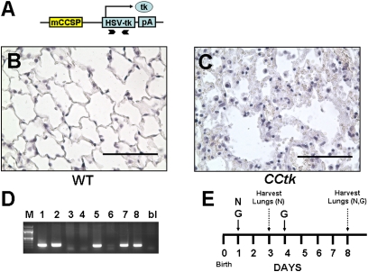
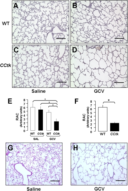
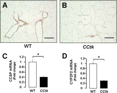
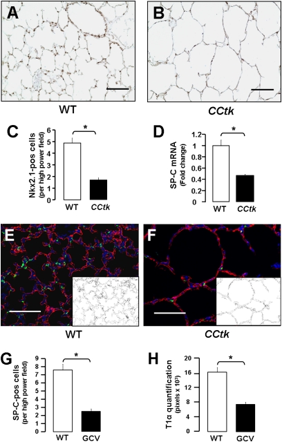
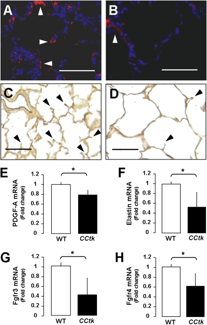
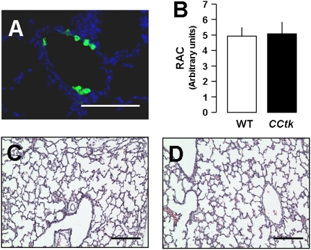
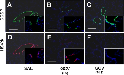
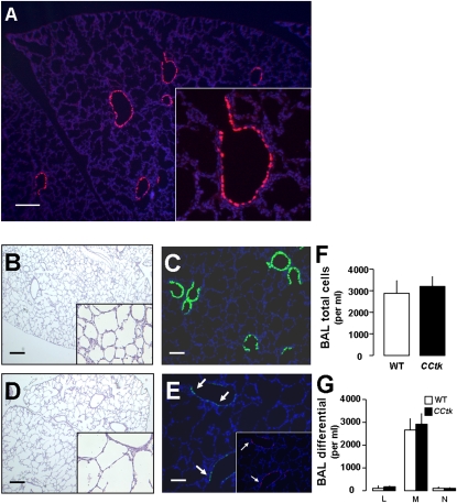
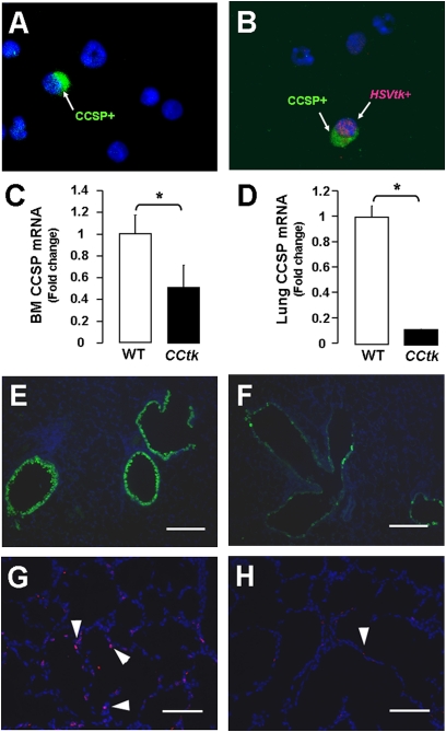
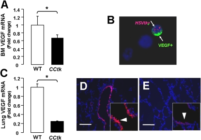
Alveolar formation is hallmarked by the transition of distal lung saccules into gas exchange units through the emergence of secondary crests and an exponential increase in surface area. Several cell types are involved in this complex process, including families of epithelial cells that differentiate into alveolar type I and II cells. Subsets of cells expressing Clara cell secretory protein (CCSP) have been identified in both lung and bone marrow compartments, and are described as a progenitor/stem cell pool involved in airway regeneration and alveolar homeostasis. Whether these cells also participate in alveolar formation during postnatal development remains unknown. Based on their regenerative capacity, we asked whether these cells participate in alveogenesis. We used a previously described transgenic mouse model (CCSP-tk) in which Ganciclovir exposure selectively depletes all cells with CCSP promoter activity through intracellular generation of a toxic metabolite of thymidine kinase. Our results showed that Ganciclovir treatment in newborn CCtk mice depleted this cell population in lung airways and bone marrow, and was associated with alveolar hypoplasia and respiratory failure. Hypoplastic lungs had fewer alveolar type I and II cells, with impaired secondary crest formation and decreased vascular endothelial growth factor expression in distal airways. These findings are consistent with a model in which a unique population of cells with CCSP promoter activity that expresses vascular endothelial growth factor participates in alveolar development.


Figures

Figure 1.
Figure 1.
Ganciclovir (GCV)-induced tk activation causes alveolar inflammation and injury in adult Clara cell secretory protein (CCSP)–herpes simplex thymidine kinase (CCtk) mice. (A) Schematic diagram of CCSP-tk transgene showing a fragment of the mouse CCSP promoter upstream of a herpes simplex virus thymidine kinase cDNA fragment flanked by bovine growth hormone polyadenylation signal. Arrow, transcription start site; chevron arrows, positions of primers for genotyping. Hematoxylin and eosin (H&E)–stained lung sections comparing GCV-treated wild-type (WT) controls animals (B) versus GCV-treated CCtk animals (C) (magnification, 40×; scale bar, 100 μM). (D) Genotyping via PCR identifying transgenic CCtk animals. (E) Schematic timeline of GCV (G) or naphthalene (N) treatment followed by lung harvest in newborn mice.
Figure 2.
Figure 2.
GCV-induced tk activation causes alveolar hypoplasia in newborn CCtk mice. H&E-stained lung sections comparing saline-treated WT control mice (A), GCV-treated WT control mice (B), saline-treated CCtk control mice (C), and GCV-treated CCtk transgenic mice (D) at 1 week after GCV treatment. Radial alveolar counts in CCtk mice compared with controls at 1 week (E) and 2 weeks (F) after GCV treatment. Representative H&E-stained lung sections in controls (G) versus CCtk mice (H) at 2 weeks after GCV treatment (magnification, 10×; scale bar, 100 μm; *P < 0.05). RAC, radial alveolar count.
Figure 3.
Figure 3.
GCV-induced tk activation depletes CCSP-positive cells in newborn CCtk mice. Immunohistochemistry for CCSP showing depleted CCSP-positive cells after GCV treatment in WT control mice (A) versus GCV-treated CCtk mice (B) (magnification, 10×; scale bar, 100 μm). Real-time quantitative PCR analysis comparing total lung mRNA after GCV treatment in CCtk mice compared with GCV-treated WT controls for CCSP mRNA expression (C) and CYP2F2 mRNA expression (D) (*P < 0.05).
Figure 4.
Figure 4.
Alveolar hypoplasia is hallmarked by fewer alveolar type I and II cells. Immunohistochemistry for Nkx2.1 after GCV treatment in WT control (A) versus GCV-treated CCtk mice (B) (magnification, 20×; scale bar, 100 μm). (C) Corresponding quantification of Nkx2.1-positive cells. (D) Real-time quantitative PCR analysis comparing total lung mRNA for surfactant protein (SP)–C mRNA expression after GCV treatment in CCtk mice compared with GCV-treated WT control animals (*P < 0.05). Immunofluorescence for SP-C and T1α after GCV treatment in control (E) versus CCtk mice (F) (magnification, 20×; scale bar, 100 μm; insets represent binary image of T1α-positive pixels). Quantitation of SP-C–positive cells (G) and T1α pixels (H) (*P < 0.05).
Figure 5.
Figure 5.
Alveolar hypoplasia is a result of impaired secondary crest formation. Immunofluorescence for α–smooth muscle actin (α-SMA) after GCV treatment in WT control (A) versus CCtk mice (B). Hart's elastin (Eln) stain after GCV treatment in WT control (C) versus CCtk mice (D). Arrowheads indicate α-SMA ([A and B]: magnification, 40×; scale bar, 100 μm) and Eln in ([C and D]: magnification, 20×; scale bar, 100 μm). Real-time quantitative PCR analysis comparing total lung mRNA after GCV treatment in CCtk mice compared with GCV-treated WT control animals for mRNA expression of platelet-derived growth factor (PDGF)–A (E), Eln (F), and fibroblast growth factor receptor (Fgfr) 3 and 4 (G and H) (*P < 0.05).
Figure 6.
Figure 6.
Naphthalene-sensitive Clara cell depletion does not impair alveolar formation. (A) Immunofluorescence for CCSP at 48 hours after naphthalene injection showing depletion of Clara cells (magnification, 40×; scale bar, 100 μm). (B) Radial alveolar counts and representative H&E-stained lung sections at 1 week after GCV treatment in WT control (C) versus CCtk mice (D) (magnification, 10×; scale bar, 100 μm).
Figure 7.
Figure 7.
GCV-induced tk activation permanently depletes tk-positive cells in newborn CCtk mouse lungs. Immunofluorescence for CCSP (AC) and HSVtk (DF) at 1 week after GCV treatment (B and E) or saline treatment (A and D) and at 2 weeks after GCV treatment (C and F) in CCtk mice (magnification, 10× [insets, 40×]; scale bar, 100 μm).
Figure 8.
Figure 8.
Potential mechanisms of GCV-induced alveolar hypoplasia. Immunofluorescence for HSVtk in 1-week-old, naive CCtk mouse lung (A). Representative lung photomicrographs of H&E-staining (B, D) and immunofluorescence (A, C, E) for CCSP (green) and HSVtk (red) lungs at 48 hours after GCV treatment in WT (B and C) versus CCtk mice (D and E), with corresponding total bronchoalveolar lavage (BAL) cells (F) and BAL differential (G) (magnification, 4× in [A–E] [insets, 40×]; scale bar, 100 μm). L, lymphocytes; M, macrophages; N, neutrophils.
Figure 9.
Figure 9.
tk-positive cells are also located in the bone marrow. Immunofluorescence from bone marrow cytospins showing CCSP-positive cells in WT mice (A) and tk/CCSP dual-positive cells in CCtk mice (B) (magnification, 40×). (C) Real-time quantitative PCR analysis after GCV treatment in CCtk mice compared with GCV-treated WT control mice for bone marrow CCSP mRNA and (D) lung CCSP mRNA expression (*P < 0.05). Immunofluorescence showing corresponding decrease in CCSP-positive cells in lungs after GCV treatment in WT control (E) versus CCtk mice (F) (magnification, 20×; scale bar, 100 μm). Ki67-positive cells (arrowheads) after GCV treatment in newborn WT control (G) versus CCtk mice (H) (magnification 20×; scale bar, 100 μm).
Figure 10.
Figure 10.
Alveolar hypoplasia is associated with decreased vascular endothelial growth factor (VEGF) expression. Real-time quantitative PCR analysis after GCV treatment in CCtk mice compared with GCV-treated WT control animals for VEGF mRNA in bone marrow (A) and lung (C) (*P < 0.05). (B) Immunofluorescence from bone marrow cytospins showing tk/VEGF dual-positive cells (arrows) in CCtk mice (magnification, 40×). Immunofluorescence of VEGF expression (arrowheads) after GCV treatment in WT control (D) versus CCtk mice (E) (magnification, 40×; scale bar, 100 μm).

Similar articles

Cited by

References

    1. Galambos C, Demello DE. Regulation of alveologenesis: clinical implications of impaired growth. Pathology 2008;40:124–140. - PubMed
    1. Husain AN, Siddiqui NH, Stocker JT. Pathology of arrested acinar development in postsurfactant bronchopulmonary dysplasia. Hum Pathol 1998;29:710–717. - PubMed
    1. Jobe AJ. The new BPD: an arrest of lung development. Pediatr Res 1999;46:641–643. - PubMed
    1. Stripp BR, Maxson K, Mera R, Singh G. Plasticity of airway cell proliferation and gene expression after acute naphthalene injury. Am J Physiol 1995;269:L791–L799. - PubMed
    1. Reynolds SD, Hong KU, Giangreco A, Mango GW, Guron C, Morimoto Y, Stripp BR. Conditional Clara cell ablation reveals a self-renewing progenitor function of pulmonary neuroendocrine cells. Am J Physiol Lung Cell Mol Physiol 2000;278:L1256–L1263. - PubMed

Publication types