. 2010 Nov 25;116(22):4512-22.
doi: 10.1182/blood-2010-02-272005. Epub 2010 Aug 6.

PPARγ activation normalizes resolution of acute sterile inflammation in murine chronic granulomatous disease

Affiliations

PPARγ activation normalizes resolution of acute sterile inflammation in murine chronic granulomatous disease

Ruby Fernandez-Boyanapalli et al. Blood. .

Abstract

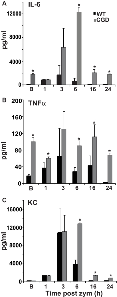
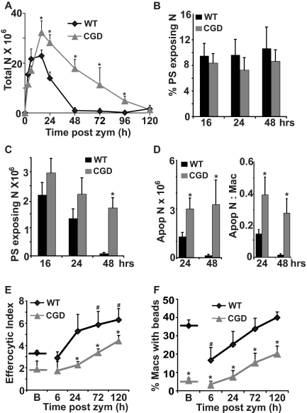
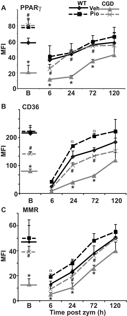
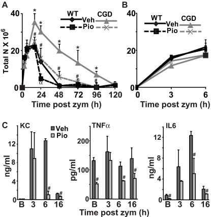
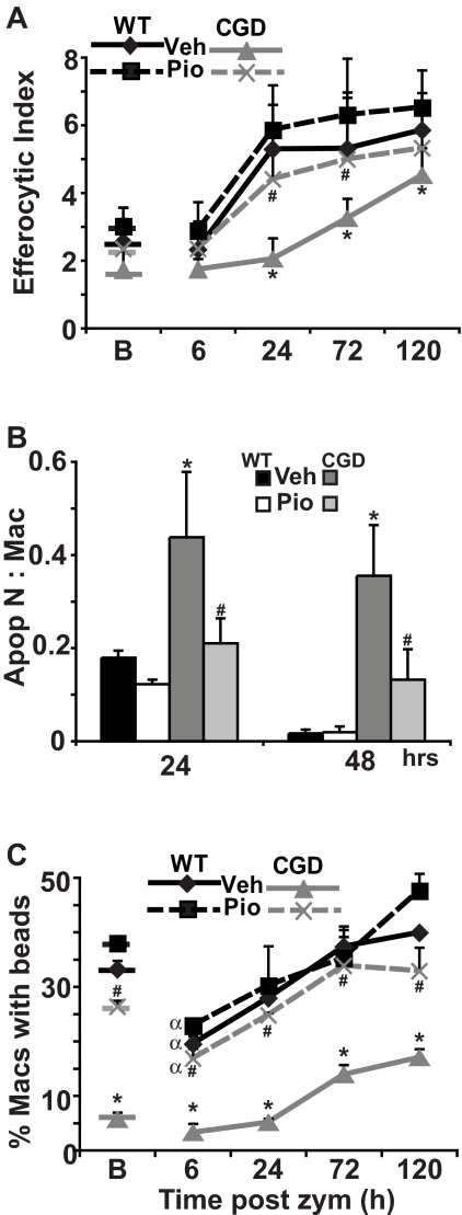
Absence of a functional nicotinamide adenine dinucleotide phosphate (NADPH) oxidase predisposes chronic granulomatous disease (CGD) patients to infection, and also to unexplained, exaggerated inflammation. The impaired recognition and removal (efferocytosis) of apoptotic neutrophils by CGD macrophages may contribute to this effect. We hypothesized that peroxisome proliferator-activated receptor γ (PPARγ) activation during CGD inflammation is deficient, leading to altered macrophage programming and decreased efferocytosis, and that PPARγ agonism would enhance resolution. using the gp91(phox-/-) murine model of X-linked CGD in a well-characterized model of sterile, zymosan-induced peritonitis, it was demonstrated that PPARγ expression and activation in CGD macrophages were significantly deficient at baseline, and acquisition was delayed over the course of inflammation relative to that of wild-type. Efferocytosis by macrophages reflected PPARγ activation during peritonitis and was impaired in CGD mice (versus wild-type), leading to accumulation of apoptotic neutrophils. Importantly, provision of the PPARγ agonist, pioglitazone, either prophylactically or during inflammation, significantly enhanced macrophage PPARγ-mediated programming and efferocytosis, reduced accumulation of apoptotic neutrophils, and normalized the course of peritonitis in CGD mice. As such, PPARγ may be a therapeutic target for CGD, and possibly other inflammatory conditions where aberrant macrophage programming and impaired efferocytosis delay resolution of inflammation.


Figures

Figure 1
Figure 1
Macrophage PPARγ expression and activity are deficient and acquisition delayed in CGD mice during peritonitis. WT and CGD mice were injected intraperitoneally with zymosan and lavage performed at the indicated times. Peritoneal F4/80 monocytes/macrophages were counted (A) and stained for intracellular PPARγ (following permeablilization) (B), surface CD36 (C), and surface macrophage mannose receptor (MMR; D). Mean fluorescence intensity (MFI) is shown (B-D). Data represent mean ± standard error (SE); N = 8 mice per time point. *P < .02 compared with WT mice at the respective time points; #P < .03 compared with baseline [B] values for each genotype, respectively.
Figure 2
Figure 2
Proinflammatory cytokines are elevated and prolonged during peritonitis in CGD mice. Peritonitis was induced as in Figure 1, and cytokines were measured in peritoneal lavage supernatants at the times indicated. [B] indicates baseline without zymosan. Data represent mean ± SE; N = 6 mice per time point investigated; *P < .03 compared with WT mice at the respective time points.
Figure 3
Figure 3
Exaggerated and prolonged neutrophilia characterizing zymosan peritonitis in CGD mice is accompanied by impaired in vivo efferocytosis by macrophages. Peritonitis was induced as in Figure 1 and peritoneal cells analyzed at the times indicated (see “Methods”). (A) Total neutrophils [N] were counted. (B) Percent of neutrophils exposing PS (annexin V–positive staining), and (C) absolute numbers of PS exposing neutrophils were determined. (D) Apoptotic neutrophils identified by nuclear morphology and the ratio of apoptotic neutrophils to macrophages were determined microscopically. (E) Efferocytic Index was determined by microscopic examination of macrophages lavaged from the inflamed peritonea. (F) Carboxylated beads were injected into the peritonea to measure in vivo efferocytic capability, and 1 hour later, the percentage of lavaged peritoneal macrophages positive for beads was determined by flow cytometry. [B] indicates baseline without zymosan. Data represent mean ± SE; N = 8 mice per time point. *P < .02 compared with WT mice at the respective time points. #P < .02 compared with WT mice at baseline.
Figure 4
Figure 4
Pioglitazone treatment enhances macrophage PPARγ expression and activity in CGD mice. Mice were gavaged with pioglitazone or vehicle for 2 days before zymosan injection and every 24 hours thereafter. F4/80-positive macrophages from lavage were analyzed as in Figure 1 for intracellular PPARγ (A), and surface CD36 (B), and MMR (C) expressed as MFI. Data represent mean ± SE; N = 8 mice per time point; #P ≤ .01 compared with vehicle-treated CGD mice at the respective time points; α,*P ≤ .01 compared with WT mice treated with vehicle, at the respective time points. [B] indicates baseline after 2 days of treatment without zymosan. Symbols for significant changes in values between baseline [B] and the early time points following zymosan for either vehicle or pioglitazone-treated mice of each genotype were as shown in Figure 1, but omitted here for simplicity.
Figure 5
Figure 5
Pioglitazone treatment enhances resolution of neutrophilia and ameliorates inflammation in CGD mice. Mice were treated as described in Figure 4. The time course of neutrophilia was determined (A, with a blowup of the early time points shown in B), and cytokines were measured in lavage supernatants (CGD mice shown) (C). Data represent mean ± SE; N = 8 mice per time point. #P ≤ .02 compared with vehicle-treated CGD mice at the respective time points, and α,*P ≤ .01 compared with WT mice treated with vehicle, at the respective time points.
Figure 6
Figure 6
Pioglitazone enhances efferocytosis and reduces accumulation of apoptotic neutrophils in CGD mice. Mice were treated as described in Figure 4. Efferocytosis by peritoneal macrophages (A) and accumulation of apoptotic neutrophils (identified by nuclear morphology) (B) were determined microscopically. In vivo uptake of carboxylated beads by macrophages was determined by flow cytometry (C) as in Figure 3. Data represent mean ± SE; N = 8 mice per time point; #P ≤ .01 compared with vehicle-treated CGD mice at the respective time points; *P ≤ .01 compared with WT mice treated with vehicle, at the respective time points. αP < .02 between baseline [B] and 6-hour after zymosan for the corresponding genotype and treatment condition, respectively.
Figure 7
Figure 7
Resolution of neutrophilia and macrophage reprogramming are enhanced by pioglitazone even when administered after onset of inflammation in CGD. Twenty-four hours after zymosan injection, mice were treated by oral gavage with a single dose of either vehicle or pioglitazone. At 48 hours after zymosan, peritonea were lavaged, and cells were analyzed as before. The course of neutrophilia is shown in panel A: solid lines represent time course of zymosan-induced peritoneal neutrophilia without treatment derived from data shown in Figure 3A; arrow shows the time at which pioglitazone or vehicle was administered; dashed lines show changes in neutrophilia following treatment. Accumulation of apoptotic neutrophils (B) and efferocytosis by macrophages (C) in peritonea at 48 hours are shown. (D) Cytokines were measured in lavage supernatants by enzyme-linked immunosorbent assay. (E) F4/80 positive macrophages were analyzed for PPARγ, CD36, and MMR by flow cytometric analysis as in Figure 1. Data represent mean ± SE; N = 8 mice per treatment group. #P ≤ .02 compared with vehicle-treated CGD mice, and *P ≤ .01 compared with vehicle-treated WT mice.

References

    1. Johnston RB., Jr Clinical aspects of chronic granulomatous disease. Curr Opin Hematol. 2001;8(1):17–22. - PubMed
    1. Rupec RA, Petropoulou T, Belohradsky BH, et al. Lupus erythematosus tumidus and chronic discoid lupus erythematosus in carriers of X-linked chronic granulomatous disease. Eur J Dermatol. 2000;10(3):184–189. - PubMed
    1. Segal BH, Leto TL, Gallin JI, Malech HL, Holland SM. Genetic, biochemical, and clinical features of chronic granulomatous disease. Medicine (Baltimore) 2000;79(3):170–200. - PubMed
    1. Holland SM. Chronic granulomatous disease. Clin Rev Allergy Immunol. 2010;38(1):3–10. - PubMed
    1. Frasch SC, Berry KZ, Fernandez-Boyanapalli R, et al. NADPH oxidase-dependent generation of lysophosphatidylserine enhances clearance of activated and dying neutrophils via G2A. J Biol Chem. 2008;283(48):33736–33749. - PMC - PubMed

Publication types

MeSH terms