. 2010 Dec;38(22):8231-8.
doi: 10.1093/nar/gkq676. Epub 2010 Aug 6.

The type II restriction endonuclease MvaI has dual specificity

Affiliations

The type II restriction endonuclease MvaI has dual specificity

Ildikó Stier et al. Nucleic Acids Res. 2010 Dec.

Abstract

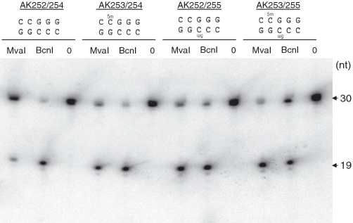
The MvaI restriction endonuclease cuts 5'-CC↓AGG-3'/5'-CC↑TGG-3' sites as indicated by the arrows. N4-methylation of the inner cytosines (C(m4)CAGG/C(m4)CTGG) protects the site against MvaI cleavage. Here, we show that MvaI nicks the G-strand of the related sequence (CCGGG/CCCGG, BcnI site) if the inner cytosines are C5-methylated: C(m5)C↓GGG/CC(m5)CGG. At M.SssI-methylated SmaI sites, where two oppositely oriented methylated BcnI sites partially overlap, double-nicking leads to double-strand cleavage (CC(m5)C↓GGG/CC(m5)C↑GGG) generating fragments with blunt ends. The double-strand cleavage rate and the stringency of substrate site recognition is lower at the methylation-dependent site than at the canonical target site. MvaI is the first restriction endonuclease shown to possess, besides the 'normal' activity on its unmethylated recognition site, also a methylation-directed activity on a different sequence.


Figures

Figure 1.
Figure 1.
(A) Digestion of SssI-methylated pUP41 DNA with MvaI and BstNI. Lanes 1, unmethylated pUP41; lanes 2, pUP41 methylated by M.SssI in vitro; lanes 3, pUP41 purified from cells expressing M.SssI; lanes 4, pUP41 purified from cells in which M.SssI production was repressed by glucose; lanes 5, pSTB–MSssI (from uninduced cells). Sizes of fragments that differ between the unmethylated and M.SssI-methylated DNAs are shown in base pairs. The band corresponding to the 543-bp fragment remains visible in the methylated sample because it also contains a comigrating 540-bp fragment. The extra fragments that appear in sub-stoichiometric amounts are indicated by asterisk. M, 1 kb DNA Ladder (Fermentas). (B) Restriction map of the plasmid pUP41. MvaI (spikes) and SmaI (arrows) sites are indicated on the perimeter.
Figure 2.
Figure 2.
MvaI cleavage at M.SssI-methylated SmaI sites. (A) Sequencing through the methylated SmaI279 site of pUP41 using intact or MvaI-cleaved templates as indicated by the scheme on the left. The terminal adenines, denoted by asterisk, are template-independent additions by Taq polymerase. (B) MvaI cleavage at the canonical recognition sequence and at the M.SssI-methylated SmaI site.
Figure 3.
Figure 3.
Comparison of MvaI cleavage rates on the canonical CCWGG/CCWGG and on the M.SssI-methylated SmaI site (CCm5CGGG/CCm5CGGG). Digestion of pUP41 plasmid DNA (∼0.5 µg) methylated in vitro by M.SssI. MvaI concentrations are shown above the lanes. M, 1 kb DNA Ladder (Fermentas); -M.SssI, unmethylated plasmid. Appearance of the 1249 and 454 bp fragments indicates cleavage of the two methylated SmaI sites present in pUP41.
Figure 4.
Figure 4.
Strand-specific nicking of M.SssI-methylated BcnI sites by MvaI. (A) Possible nicking mechanisms at M.SssI-methylated BcnI sites. (B) Sequencing through the M.SssI-methylated and MvaI-digested BcnI2411 site of pUP41. Asterisk, template-independent addition by the Taq polymerase.
Figure 5.
Figure 5.
MvaI digestion of oligonucleotides containing unmethylated, hemimethylated or fully methylated BcnI sites. Electrophoresis of cleavage products in a 10% denaturing polyacrylamide gel. The 30-mer oligonucleotide duplexes contained unmethylated (AK252/254), hemimethylated (AK253/254 and AK252/255) or fully methylated (AK253/255) BcnI sites as shown above the gel. In all duplexes, the G-strand (the strand with G in the central position) was radioactively labeled at the 3′-end. Appearance of a 19-nt fragment indicates cleavage of the G-strand.
Figure 6.
Figure 6.
MvaI digestion of the 32P-labeled 444 bp pUC18 DNA fragment containing an unmethylated BcnI site. (A) Electrophoresis in a 6% denaturing polyacrylamide gel. Lane 1, undigested; lanes 2 and 5, digested with BcnI; lanes 3 and 6, digested with 0.5 U/µl MvaI; lanes 4 and 7, digested with 1.0 U/µl MvaI. Samples in lanes 5, 6 and 7 also contained pUC18 DNA as internal control to test completeness of digestion. Nicking of the G-strand or double-strand cleavage at the single BcnI site produces a 175-nt single-stranded labeled fragment. (B) ImageQuant line graph of lanes 1, 2 and 4. (C) Agarose gel electrophoresis of aliquots of the samples in lanes 5, 6 and 7. pUC, pUC18 completely digested with BcnI or MvaI. M, 1 kb DNA Ladder (Fermentas).

Similar articles

Cited by

References

    1. Roberts RJ, Belfort M, Bestor T, Bhagwat AS, Bickle TA, Bitinaite J, Blumenthal RM, Degtyarev S, Dryden DT, Dybvig K, et al. A nomenclature for restriction enzymes, DNA methyltransferases, homing endonucleases and their genes. Nucleic Acids Res. 2003;31:1805–1812. - PMC - PubMed
    1. Roberts RJ. How restriction enzymes became the workhorses of molecular biology. Proc. Natl Acad. Sci. USA. 2005;102:5905–5908. - PMC - PubMed
    1. Pingoud A, Jeltsch A. Structure and function of type II restriction endonucleases. Nucleic Acids Res. 2001;29:3705–3727. - PMC - PubMed
    1. Gowers DM, Bellamy SR, Halford SE. One recognition sequence, seven restriction enzymes, five reaction mechanisms. Nucleic Acids Res. 2004;32:3469–3479. - PMC - PubMed
    1. Smith HO, Wilcox KW. A restriction enzyme from Hemophilus influenzae. I. Purification and general properties. J. Mol. Biol. 1970;51:379–391. - PubMed

Publication types

MeSH terms

Substances