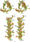
Structure of the N-terminal fragment of Escherichia coli Lon protease
- PMID: 20693685
- PMCID: PMC2917273
- DOI: 10.1107/S0907444910019554
Structure of the N-terminal fragment of Escherichia coli Lon protease
Abstract
The structure of a recombinant construct consisting of residues 1-245 of Escherichia coli Lon protease, the prototypical member of the A-type Lon family, is reported. This construct encompasses all or most of the N-terminal domain of the enzyme. The structure was solved by SeMet SAD to 2.6 A resolution utilizing trigonal crystals that contained one molecule in the asymmetric unit. The molecule consists of two compact subdomains and a very long C-terminal alpha-helix. The structure of the first subdomain (residues 1-117), which consists mostly of beta-strands, is similar to that of the shorter fragment previously expressed and crystallized, whereas the second subdomain is almost entirely helical. The fold and spatial relationship of the two subdomains, with the exception of the C-terminal helix, closely resemble the structure of BPP1347, a 203-amino-acid protein of unknown function from Bordetella parapertussis, and more distantly several other proteins. It was not possible to refine the structure to satisfactory convergence; however, since almost all of the Se atoms could be located on the basis of their anomalous scattering the correctness of the overall structure is not in question. The structure reported here was also compared with the structures of the putative substrate-binding domains of several proteins, showing topological similarities that should help in defining the binding sites used by Lon substrates.
Figures


Similar articles
-
Crystal structure of the N-terminal domain of E. coli Lon protease.Protein Sci. 2005 Nov;14(11):2895-900. doi: 10.1110/ps.051736805. Epub 2005 Sep 30. Protein Sci. 2005. PMID: 16199667 Free PMC article.
-
Crystal structure of the AAA+ alpha domain of E. coli Lon protease at 1.9A resolution.J Struct Biol. 2004 Apr-May;146(1-2):113-22. doi: 10.1016/j.jsb.2003.09.003. J Struct Biol. 2004. PMID: 15037242 Review.
-
Crystal structure of the N domain of Lon protease from Mycobacterium avium complex.Protein Sci. 2019 Sep;28(9):1720-1726. doi: 10.1002/pro.3687. Protein Sci. 2019. PMID: 31306520 Free PMC article.
-
The active site of a lon protease from Methanococcus jannaschii distinctly differs from the canonical catalytic Dyad of Lon proteases.J Biol Chem. 2004 Dec 17;279(51):53451-7. doi: 10.1074/jbc.M410437200. Epub 2004 Sep 28. J Biol Chem. 2004. PMID: 15456757
-
Slicing a protease: structural features of the ATP-dependent Lon proteases gleaned from investigations of isolated domains.Protein Sci. 2006 Aug;15(8):1815-28. doi: 10.1110/ps.052069306. Protein Sci. 2006. PMID: 16877706 Free PMC article. Review.
Cited by
-
Functional characterization of LotP from Liberibacter asiaticus.Microb Biotechnol. 2017 May;10(3):642-656. doi: 10.1111/1751-7915.12706. Epub 2017 Apr 5. Microb Biotechnol. 2017. PMID: 28378385 Free PMC article.
-
Complete three-dimensional structures of the Lon protease translocating a protein substrate.Sci Adv. 2021 Oct 15;7(42):eabj7835. doi: 10.1126/sciadv.abj7835. Epub 2021 Oct 15. Sci Adv. 2021. PMID: 34652947 Free PMC article.
-
Defining the crucial domain and amino acid residues in bacterial Lon protease for DNA binding and processing of DNA-interacting substrates.J Biol Chem. 2017 May 5;292(18):7507-7518. doi: 10.1074/jbc.M116.766709. Epub 2017 Mar 14. J Biol Chem. 2017. PMID: 28292931 Free PMC article.
-
Structure and the Mode of Activity of Lon Proteases from Diverse Organisms.J Mol Biol. 2022 Apr 15;434(7):167504. doi: 10.1016/j.jmb.2022.167504. Epub 2022 Feb 17. J Mol Biol. 2022. PMID: 35183556 Free PMC article. Review.
-
Roles of the N domain of the AAA+ Lon protease in substrate recognition, allosteric regulation and chaperone activity.Mol Microbiol. 2014 Jan;91(1):66-78. doi: 10.1111/mmi.12444. Epub 2013 Nov 10. Mol Microbiol. 2014. PMID: 24205897 Free PMC article.
References
-
- Amerik, A. Y., Antonov, V. K., Ostroumova, N. I., Rotanova, T. V. & Chistyakova, L. G. (1990). Bioorgan. Khim. 16, 869–880. - PubMed
-
- Bolon, D. N., Wah, D. A., Hersch, G. L., Baker, T. A. & Sauer, R. T. (2004). Mol. Cell, 13, 443–449. - PubMed
-
- Botos, I., Melnikov, E. E., Cherry, S., Khalatova, A. G., Rasulova, F. S., Tropea, J. E., Maurizi, M. R., Rotanova, T. V., Gustchina, A. & Wlodawer, A. (2004). J. Struct. Biol. 146, 113–122. - PubMed
-
- Botos, I., Melnikov, E. E., Cherry, S., Kozlov, S., Makhovskaya, O. V., Tropea, J. E., Gustchina, A., Rotanova, T. V. & Wlodawer, A. (2005). J. Mol. Biol. 351, 144–157. - PubMed
-
- Botos, I., Melnikov, E. E., Cherry, S., Tropea, J. E., Khalatova, A. G., Rasulova, F., Dauter, Z., Maurizi, M. R., Rotanova, T. V., Wlodawer, A. & Gustchina, A. (2004). J. Biol. Chem. 279, 8140–8148. - PubMed
Publication types
MeSH terms
Substances
Associated data
- Actions
Grants and funding
LinkOut - more resources
Full Text Sources
Other Literature Sources
Molecular Biology Databases

