Review
. 2010 Jul-Aug;16(4):382-91.
doi: 10.1097/PPO.0b013e3181eaca65.

Toll-like receptor agonists: are they good adjuvants?

Affiliations
Review

Toll-like receptor agonists: are they good adjuvants?

Sacha Gnjatic et al. Cancer J. 2010 Jul-Aug.

Abstract

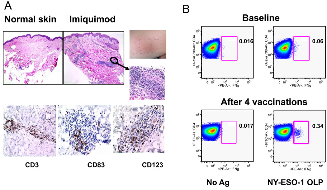
Therapeutic immunization leading to cancer regression remains a significant challenge. Successful immunization requires activation of adaptive immunity, including tumor specific CD4 T cells and CD8 T cells. Generally, the activation of T cells is compromised in patients with cancer because of immune suppression, loss of tumor antigen expression, and dysfunction of antigen-presenting cells. Antigen-presenting cells such as dendritic cells (DCs) are key for the induction of adaptive antitumor immune responses. Recently, attention has focused on novel adjuvants that enhance dendritic cell function and their ability to prime T cells. Agonists that target toll-like receptors are being used clinically either alone or in combination with tumor antigens and showing initial success both in terms of enhancing immune responses and eliciting antitumor activity. This review summarizes the application of these adjuvants to treat cancer and the potential for boosting responses in vivo.


Conflict of interest statement

Conflict of Interest Statement:

All authors have no conflict of interest to declare.

Figures

Figure 1
Figure 1. Expression of Toll-like receptors on innate immune cells
TLR 1,-2,-4, -5 and -6 are expressed in the plasma membrane where TLR2 associates with either TLR1 or TLR6. TLR3, -7, -8 and -9 traffic from the endoplasmic reticulum to the endosome where they encounter their ligands. MYD88 (myeloid differentiation primary response protein 88) and TRIF (TIR domain-containing adaptor protein inducing IFN) are signalling adaptors that link Toll-like receptors (TLRs) are to downstream kinases that define a given signalling pathway. All TLRs use MyD88, except for TLR3 which uses TRIF. The sorting adaptor TIRAP (TIR domain-containing adaptor protein) is used by TLR1, TLR2, TLR4 and TLR6 and links the TIR domain to MyD88, whereas TRIF is recruited by both TLR4 and TLR3. An additional adaptor TRAM, links the TIR domain of TLR4 with TRIF. TLRs which use the MyD88 dependent pathway recruit the IRAK family of proteins and TRAF6 resulting in the activation of TAK1. This in turn leads to the activation of NFKB and the MAPK pathway and results in the induction of pro-inflammatory cyokines and upregulation of phenotypic markers of activation (CD80, CD86). TLR4 (which relies on additional accessory molecules MD2 and CD14) and TLR3 both trigger the TRIF-dependent pathway, which also leads to activation of inflammatory cytokines via NFKB and MAP Kinase. In addition, TRIF recruits TRAF, leading to the activation of TBK1/IKKi, IRF3 and IRF7 and transcription of type I IFN. MyD88 also associates with the IRAK family of proteins. A complex of proteins (TRAF3, IRAK1 and Ikkα) subsequently activates IRF7. Examples of ligands binding the TLRs are shown. (Adapted from Kumar et al.,(125)). Abbreviations: LPS, lipopolysaccharide; PtdIns(4,5)P2, phosphatidylinositol-4,5-bisphospate; TRAF3, TNFR-associated factor 3.
Figure 2
Figure 2. Major DC subsets in blood
There are two subsets in blood, the myeloid DC (mDC) or the plasmacytoid DC (pDC). They are distinguished by surface marker expression, TLR expression, cytokine production and primary functional roles. It is now appreciated that pDC can also participate in the induction of adaptive immune responses although their precise roles need to be determined. While pDC do not synthesize IL-12, mDC can produce type I IFN via TLR3 ligation.
Figure 3
Figure 3. DC undergo activation following ligation of TLR and prime CD4+ and CD8+ T cells
A. DC are most efficient at acquiring antigen when they are in their immature state through mechanisms that include phangocytosis, endocytosis and receptor mediated uptake. After encountering TLR ligands, they undergo maturation and upregulate HLA molecules (which present peptide antigens to T cell receptors on T cells) as well as co-stimulatory molecules such as CD80 and CD86, which interact with CD28 on T cells. DC also produce cytokines (IL-12, type I IFN) that aid in priming of CD4+ helper cells and cytolytic T cells. B. DC utilize endogenous and exogenous pathways to process and present antigens to CD8+ T cells. In the endogenous pathway, exemplified by virus infection or transduction of cells with RNA or DNA encoding antigens, antigen is processed in the cytoplasm by the proteosome and then transported into the ER where further processing can take place and peptides access newly synthesized HLA class I molecules. The peptide-HLA complex is then transported to the cell surface where it can interact with the T cell receptor. In the exogenous pathway, dying virus- infected cells or tumor cells (e.g. following chemotherapy or irradiation) are phagocytosed by DC and crosspresented to T cells. Dying tumor cells also release factors that activate DC via TLRs or components of the inflammasome. Antigens from these cells may access the cytoplasm and intersect with the conventional endogenous pathway of antigen processing. Alternatively, they may be processed within the endosomes themselves and acquired by recycling class I molecules which return to the cell surface. The exogenous pathway explains how antigens from dead cells can be acquired and presented to T cells.
Figure 4
Figure 4. Imiquimod induces local inflammation and NY-ESO-1 specific CD4+ T cell responses
A. Representative H and E stained sections of control skin and Imiquimod treated skin (left panels). Right upper panels show inflammation at the Imiquimod treated of one patient. Representative immunohistochemistry sections for three tested markers are shown (CD3: T cells; CD83: mature DC; CD123: plasmacytoid DC). B. Quantification of IFNγ-secreting NY-ESO-1-specfic CD4+ T cells. Representative before and after vaccine samples for one patient are shown. Following a one week in vitro stimulation with pooled NY-ESO-1 overlapping peptides, cells were re-stimulated and stained for intracellular IFNγ. CD4 staining is shown on the y axis and IFNγ staining is shown on the x-axis.(76). Copyright 2008. The American Association of Immunologists, Inc.

References

    1. Smyth MJ, Dunn GP, Schreiber RD. Cancer immunosurveillance and immunoediting: the roles of immunity in suppressing tumor development and shaping tumor immunogenicity. Advances in immunology. 2006;90:1–50. - PubMed
    1. Anderson KV, Bokla L, Nusslein-Volhard C. Establishment of dorsal-ventral polarity in the Drosophila embryo: the induction of polarity by the Toll gene product. Cell. 1985;42:791–798. - PubMed
    1. Lemaitre B, Reichhart JM, Hoffmann JA. Drosophila host defense: differential induction of antimicrobial peptide genes after infection by various classes of microorganisms. Proceedings of the National Academy of Sciences of the United States of America. 1997;94:14614–14619. - PMC - PubMed
    1. Pichlmair A, Reis e Sousa C. Innate recognition of viruses. Immunity. 2007;27:370–383. - PubMed
    1. Kawai T, Akira S. Innate immune recognition of viral infection. Nature immunology. 2006;7:131–137. - PubMed

Publication types

MeSH terms