

Cochlear otosclerosis
- PMID: 20693902
- PMCID: PMC3075959
- DOI: 10.1097/MOO.0b013e32833d11d9
Cochlear otosclerosis
Abstract
Purpose of review: The aim of this study is to summarize current advances in research and clinical aspects of cochlear otosclerosis.
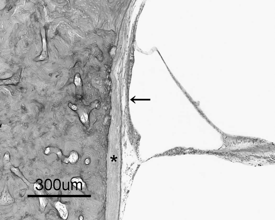
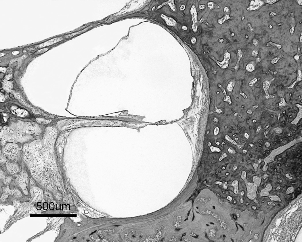
Recent findings: Recent studies have revealed that otosclerosis is a process of bone remodeling that is unique to the otic capsule only. Even though no obvious bone remodeling is seen in the otic capsule under normal conditions, remodeling starts when some molecular factors trigger the capsule in certain patients who have genetic and/or environmental tendencies.
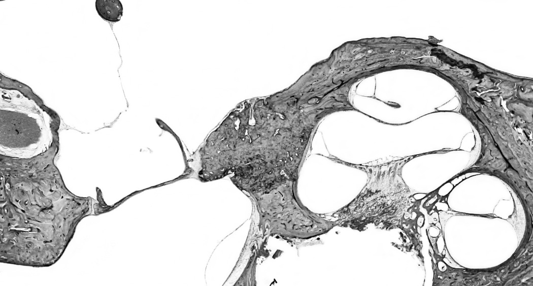
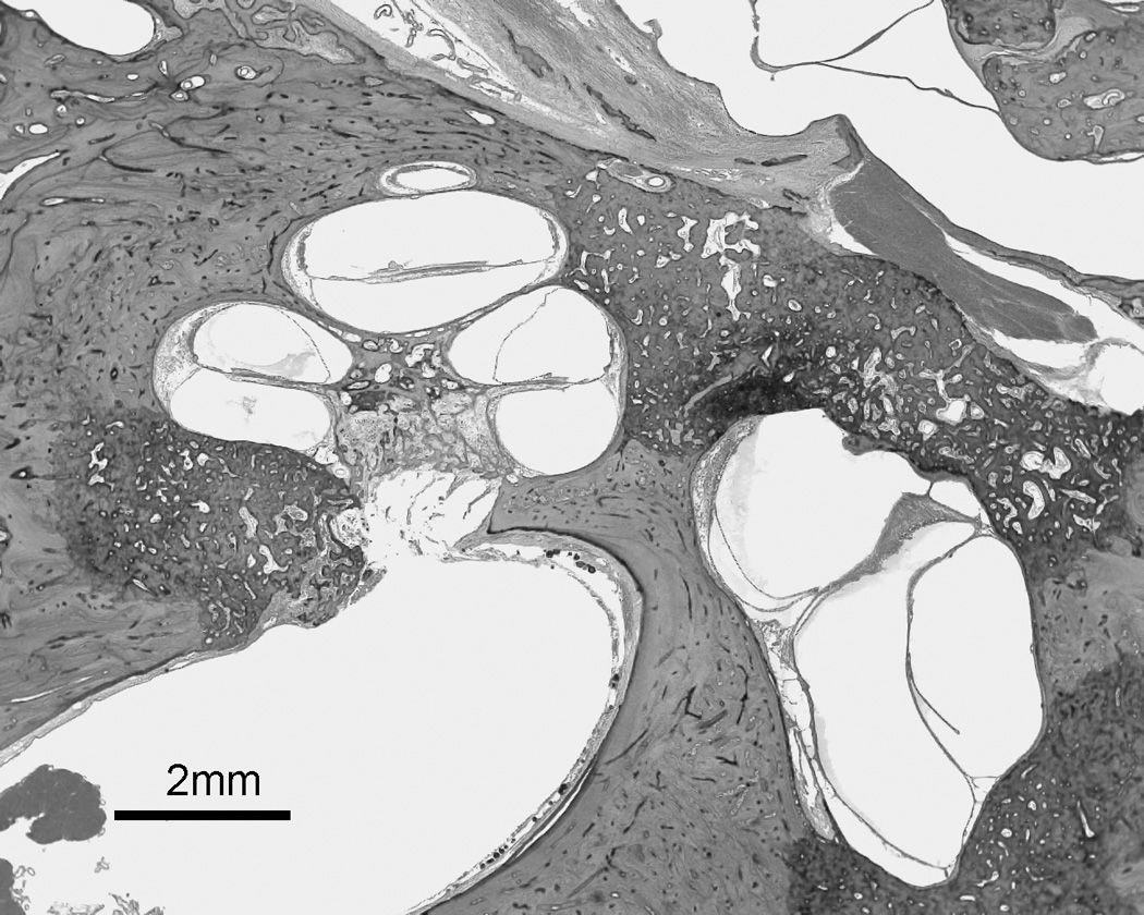
Summary: Cochlear otosclerosis is defined as otosclerosis located in the otic capsule involving the cochlear endosteum and causing sensorineural hearing loss or mixed-type hearing loss. It has been clearly shown that, when otosclerosis is sufficiently severe to involve the cochlear endosteum, it usually fixes the stapes as well.
Figures
References
-
- Schuknecht HF, Kirchner JC. Cochlear otosclerosis: fact or fantasy. Laryngoscope. 1974;84:766–782. - PubMed
-
- Frisch T, Sørensen MS, Overgaard S, Bretlau P. Estimation of volume referent bone turnover in the otic capsule after sequential point labeling. Ann Otol Rhinol Laryngol. 2000;109:33–39. - PubMed
-
- McKenna MJ, Kristiansen AG. Molecular biology of otosclerosis. Adv Otorhinolaryngol. 2007;65:68–74. - PubMed
-
- Tomek MS, Brown MR, Mani SR, et al. Localization of a gene for otosclerosis to chromosome 15q25–q26. Hum Mol Genet. 1998;7:285–290. - PubMed
Publication types
MeSH terms
Grants and funding
LinkOut - more resources
Full Text Sources
Research Materials